


SERS: High-Tech Tactic May Newly Expose Stealthy Salmonella
USDA ARS researcher, Dr Bosoon Park, and his team have been evaluating the strengths and shortcomings of ‘surface–enhanced Raman scattering’ (SERS) for Salmonella testing compared to common analytical methods and several newer ones as well, with the view to gaining better control over the world’s worst foodborne pathogens. 29 May 2012
 6 minute read
At laboratories of the future, even the smallest quantity of Salmonella bacteria may be easily detected with a technology known as ‘SERS’, short for ‘surface-enhanced Raman scattering’.
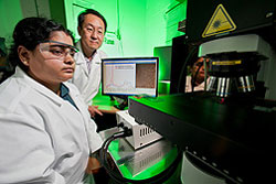
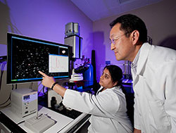
Agricultural engineer, Dr Bosoon Park, in the Agricultural Research Service’s Quality and Safety Assessment Research Unit in Athens, Georgia, is leading exploratory studies of this analytical technique’s potential for quick, easy and reliable detection of Salmonella and other foodborne pathogens.
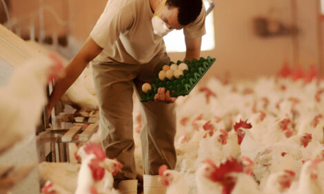
According to the US Centers for Disease Control and Prevention, Salmonella causes more than one million cases of illness in the US every year.
If SERS proves successful for cornering Salmonella, the technique might be used at public health laboratories around the nation to rapidly identify pathogens responsible for outbreaks of foodborne illness. Tomorrow’s food–makers might opt to use SERS at their in-house quality-control labs to help ensure that their products are free of unsafe levels of this or other harmful bacteria.
For Salmonella testing, labs have many choices. They can opt for well-established technologies, such as monoclonal antibody assays, ELISA (enzyme-linked immunosorbent assay) or PCR (polymerase chain reaction) tests.
Dr Park’s team is evaluating the strengths and shortcomings of SERS in relation to these popular analytical methods and several newer ones as well, including atomic force microscopy and surface plasmon resonance.
According to Dr Park, a SERS analysis is relatively simple to perform, an advantage when labs are swamped with samples from an outbreak of foodborne illness or a product recall.
With further research, SERS may offer superior detection capabilities, making it faster and easier to find very small quantities of bacteria in a complex, real-world background, such as a food or beverage sample. That is especially important, since it is apparently possible for as few as 10 Salmonella cells to cause illness, depending on the strain involved and other factors.
Nobel Laureate’s Work is Basis of Today’s SERS
Developed over the past two decades or so, SERS has expanded the power and potential of Raman spectroscopy, its parent technology. Raman spectroscopy is based on discoveries made in the early 1900s by Sir Chandrasekhara V. Raman, who received the 1930 Nobel Prize in physics for his research.
In a SERS analysis, a specimen is placed on a surface, such as a stainless steel plate, silicon wafer, or glass slide, that has been ‘enhanced’ – or changed from smooth to rough. For example, a glass slide or silicon wafer could be etched, to roughen it, or a small stainless steel plate could be coated with an extremely thin ‘nanolayer’ of minuscule particles of a metal, such as silver, gold or copper.
In some of its research, Dr Park’s team enhanced the surface of stainless steel plates by coating them with tiny spheres, made up of a biopolymer encapsulated with silver nanoparticles.
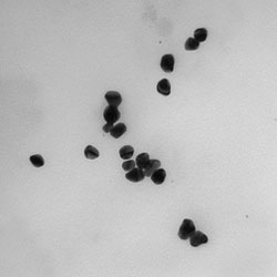
Rough surfaces and colloidal metals, such as silver, can enhance the scattering of light that occurs when a specimen, placed on this ‘nanosubstrate’, is scanned with the Raman spectrometer’s laser beam. The scattered light that comes back to the spectroscope forms a distinct spectral pattern known as a ‘Raman spectral signature’, or a ‘Raman scattered signal’.
“The idea of using a substrate of silver nanoparticles for Raman spectroscopy is not new,” says Dr Park. “But in SERS studies to detect foodborne pathogens, our use of a substrate of silver nanoparticles that encapsulate a biopolymer is novel, to the best of our knowledge.”
Though SERS research with biological specimens is still in comparatively early stages, researchers expect to prove the concept that all molecules, such as those that make up Salmonella, have their own unique Raman spectral signature. In the future, the Raman signature of an unidentified biological specimen could be compared to known Raman signatures to find a match and thus identify the unknown specimen.
Dr Park’s team has already developed preliminary Raman spectral signatures for two common pathogenic kinds, or serotypes, of S. enterica – Enteritidis and Typhimurium – collected from raw chicken.
Silver-Encapsulated Spheres Tested as a Nanosubstrate
For this research, the group used a biopolymer encapsulated with silver as the nanosubstrate, and they worked with comparatively large concentrations of the serotypes (about 108 colony-forming units per millilitre of solution; cfu per ml). The high concentrations simplified detection and showed, apparently for the first time, that SERS can differentiate the two Salmonella serotypes by their Raman signatures.
Plans call for developing a database, or library, of Raman spectral signatures of these Salmonella serotypes and other major foodborne pathogens at various concentrations. Ideally, the signatures would be posted on the Internet for public access by researchers and analytical labs worldwide.
In follow-up studies, Dr Park intends to use less-concentrated samples of Salmonella serotypes to challenge more rigorously the technology’s ability to detect and quantify the pathogen. These tests may ultimately reveal SERS’s sensitivity, or detection limit – the smallest number of Salmonella cells, in a given concentration of solution, that can be accurately and reliably detected with a given combination of variables. Those variables might include the types of SERS instruments, sample preparation method, sample nanosubstrate, power and duration of the laser beam scanning, and more.
In an earlier experiment, using a different silver-based substrate, the team showed that SERS can differentiate live Salmonella cells from dead ones. That is significant. It could help quality-control labs evaluate the performance of today’s food-sterilisation methods, such as high-pressure processing, irradiation and thermal processing. In addition, reliable differentiation can speed and simplify the work of researchers who are developing and testing new techniques to keep food safe to eat.
“SERS is one of several candidate technologies we are currently testing,” says Dr Park. “If we find that it continues to offer major advantages over other options, our goal would be to develop a SERS-based system that wouldn’t require extensive training and would use affordable SERS instruments.”
Dr Park has presented some of these findings at an SPIE (Society of Photo-Optical Instrumentation Engineers) conference in Orlando, Florida. His collaborators include Yao-Wen Huang and Yiping Zhao of the University of Georgia-Athens; Arthur Hinton, Jr., Kurt C. Lawrence, Jaya Sundaram, William R. Windham, and Seung Chul Yoon, all with ARS at Athens; and Yongkuk Kwon of South Korea’s Animal, Plant, and Fisheries Quarantine and Inspection Agency.
The team’s studies may do much to keep us safe from some of the world’s worst foodborne pathogens.
 17 April 2024
8
minute read
17 April 2024
8
minute read