


VLA: Spotty Liver Disease in Free-Range Poultry
UX - An outbreak of Spotty Liver Disease is among the cases reported in the October 2009 VLA Monthly Scanning Surveillance Report. 14 January 2010
 5 minute read
Commercial Layers and Layer Breeders
Vibrionic hepatitis ('Spotty Liver Disease')
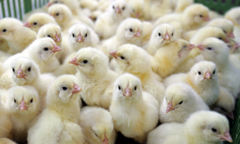
The carcasses of five free-range laying hens aged approximately 25-weeks were submitted to Winchester as part of an investigation into increased mortality. Post mortem examination revealed the presence in four out of the five carcasses of enlarged livers, which were firm to the touch. Multiple miliary 1 to 2mm diameter pale lesions were present throughout the entire liver substance. The gross post mortem findings are suggestive of 'spotty liver disease', a recognised cause of mortality within free-range units. Campylobacter jejuni was isolated from liver and splenic tissue with no other significant microbiological findings. Histopathology confirmed the presence of a multifocal fibrinoid hepatic necrosis together with a fibrinous splenitis. A similar clinical picture was reported in the Veterinary Record (2003), 153: 664.
Fowl cholera
Fowl cholera was found to be the cause of a sudden increase of mortality in a flock of 64-week-old free-range layer hens. Pasteurella multocida was recovered in pure cultures from typical lesions of septicaemia and acute egg yolk peritonitis.
Colisepticaemia
E. coli septicaemia and egg peritonitis was confirmed as the cause of increased mortality in two flocks of free-range layers aged 24 and 58 weeks, respectively. Post-mortem examination revealed septicaemic carcasses with swollen livers and spleens, and egg peritonitis. Profuse pure growth of E. coli was cultured from spleen liver and peritoneum. In addition, pecking damage to the flank and tail was seen in the older flock.
Broilers and Broiler Breeders
Rickets
Rickets was diagnosed in two flocks of 21- and 22-day-old broilers with a history of poor growth, recent deterioration of litter condition and recent onset of lameness. Findings at post mortem examination were of poor bone strength affecting leg bones in particular, and slight enlargement of parathyroid glands. Histological examination of undecalcified sections of growth plates stained by Von Kossa's method confirmed mild osteodystrophic changes and patchy mineralisation failure consistent with rickets.
Mycotic pneumonia
Lungs (threes sets) from 26-day-old broilers were submitted to Preston to investigate a problem of increasing mortality (1.8 per cent overall in a group of 36,000) and respiratory disease. Multiple pale white/cream foci up to 2mm diameter were found throughout the lungs. Branching septate fungal hyphae were found on direct microscopy if the lesions, which yielded Aspergillus fumigatus and Aspergillus versicolor on culture – both potential pathogens. Histopathology confirmed the diagnosis of chronic severe multifocal granulomatous mycotic pneumonia.
Turkeys
Blackhead and mycoplasmosis
Post mortem examination of a 12-week-old turkey at Bury revealed thickening of the air sacs with perihepatitis and pericarditis in a 12-week-old turkey. There were white firm masses in the liver, parenchyma and some thickening of the caecal wall. On histology, the liver lesions were shown to be consistent with chronic histomoniasis (although the organism was no longer detectable). Mycoplasma synoviae was detected in this bird by DGGE PCR examinations of lung tissue and may have been contributing to the airsacculitis whereas E. coli was suspected as a secondary contributor to a septicaemia.
Rickets
A group of 80 turkeys had ten birds start to go off their legs at three weeks old. Three severely affected birds were submitted to Newcastle for post mortem examination. They had been purchased at day-old and fed proprietary concentrate feed. Histopathology revealed osteodystrophy and mineralisation failure consistent with rickets. The pattern of changes suggested a 'hypophosphataemic' rickets.
Candidiasis
Crop candidiasis was seen in two flocks of 1500, 49-day-old and 600, eight-week-old turkeys with a history of poor growth and slight increase in mortality. Post mortem examination revealed focal to extensive pseudo-membranous formation of the crop mucosa and marked bursal atrophy. Histological examination confirmed the presence of PAS positive yeast pseudohyphae and a severe lymphoid depletion suggestive of immunosuppression.
Gamebirds
Staphylococcus aureus septicaemia caused the death of a 14-week-old red-legged partridge. Five red-legged partridges had died suddenly out of a group of 80 in the two weeks since purchase. The birds were generally in good condition. A pure growth of Staphylococcus aureus was isolated from liver and lung and in mixed growth from spleen.
Further Reading
| - | You can view the full report by clicking here. |
Further Reading
| - | Find out more information on the diseases mentioned in this article by clicking here. |