


VLA: Blackhead, Mycoplasmosis in Turkeys
UK - Blackhead and mycoplasmosis caused losses in turkeys, according to the Veterinary Laboratories Agency (VLA) Monthly Scanning Surveillance Report for October 2010. 16 February 2011
 5 minute read
Commercial Layers and Layer Breeders
Fowl cholera
Aberystwyth isolated Pasteurella multocida in septicaemic distribution in three, Lohmann Brown free-range chickens from a group of 6,000 aged 63 weeks. Daily death rate had increased from about three to between seven and ten. No clinical signs were seen in the other birds. Egg production was as expected for age, and there had been no change in egg quality except for an increase in white eggs. The main finding at necropsy was 'egg peritonitis'.
Pasteurella multocida isolated from all sites cultured showed resistance to tetracycline.
Enterococcus hirae
Increased mortality in two houses of five-day-old pullet chicks was attributed to a septicaemia due to Enterococcus hirae and E. coli. Post mortem findings included swollen livers and enlarged and pale spleens. Routine bacteriological cultures of livers and spleens yielded heavy virtually pure growths of Enterococcus species with biochemical features consistent with Enterococcus hirae in two samples and heavy growth of E. coli in a further sample.
Turkeys
Blackhead
Intermittent losses totalling 30 of 150 free-range turkeys during the 10 weeks after placement at day-old was found to be caused by blackhead following necropsy at Luddington of two birds that had presented with malaise and pale yellow faeces prior to death. Typical blackhead lesions were seen in the liver and one bird showed extensive thickening and necrosis of the caecal mucosal wall.
Bury and Langford also diagnosed the condition, Langford reporting three cases affecting 12- to 14-week old turkeys dying shortly after becoming listless and developing yellow/green diarrhoea.
Mycoplasmosis
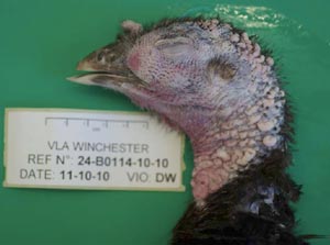
Winchester investigated a problem of nasal discharge, facial swelling and sneezing affecting twelve birds out of a flock of 144 turkeys aged 12 to 14 weeks. Post-mortem examination revealed marked swelling of the infra-orbital sinuses (see figure). Initial investigations failed to confirm the involvement of Mycoplasma gallisepticum or TRT but the use of a more sensitive serological test subsequently showed that both birds were positive for Mycoplasma synoviae and M. meleagridis, and one was positive for M. gallisepticum.
Broilers and Broiler Breeders
Rickets
Rickets was diagnosed in a flock of 31-day-old broilers with a history of poor growth. Findings at post mortem examination were of poor bone strength affecting leg bones and slight enlargement of parathyroid glands. Histological examination of undecalcified sections of growth plates stained by Von Kossa's method confirmed osteodystrophic changes and mineralisation failure consistent with hypocalcaemic rickets.
Brachyspira
A problem of loose faeces affecting approximately 20 per cent of a group of 30-week-old free-range laying hens was investigated. Culture of faeces revealed Brachyspira intermedia in all three of the houses sampled. This organism has been isolated from birds with loose faeces but the exact aetiology of spirochaetal diarrhoea is not clear.
Backyard Flocks
Infectious laryngotracheitis
Infectious laryngotracheitis (ILT) was suspected in tracheal samples showing an acute haemorrhagic/ulcerative tracheitis with inclusion bodies, from a flock of 400 birds from which 50 had died. There was a history of coughing, gasping and sinusitis in the birds.
Mycoplasmosis
Infectious sinusitis was diagnosed in an 18-week-old pullet from a flock of 60, showing respiratory signs and recent onset of lameness. The bird was emaciated, lethargic and unresponsive. Findings at post mortem examination, although mostly unremarkable, included small amount of clear mucus in the left sinus and mid trachea. Histological examination of trachea and nasal passages revealed lesions suggestive of mycoplasma involvement. Serology for antibodies to Mycoplasma gallisepticum was strongly positive confirming the diagnosis.
Ducks and Geese
Yersiniosis
A chronic bacterial infection with pneumonia and air sacculitis due to Yersinia pseudotuberculosis infection was seen in a small flock of 30-day-old Barbary ducks submitted with a history of increased mortality. Post mortem examination revealed marked congestion and consolidation of the lungs and thoracic airsacculitis. Histological examination of lungs revealed a multifocal fibrinogranulocytic and granulomatous pneumonia with numerous bacterial colonies. Appropriate advice on control was given including the zoonotic aspects.
Further Reading
| - | You can view the full report by clicking here. |
Further Reading
| - | Find out more information on the diseases mentioned in this article by clicking here. |