


AHVLA Scanning Surveillance Report - August 2011
UK - Spotty Liver syndrome was detected in a 28-week-old layer flock. 4 November 2011
 6 minute read
Commercial Layers and Layer Breeders
Caecal coccidiosis in layer pullets
Twenty-eight of 350, 16-day-old layer pullet chicks died in two days. Postmortem examination showed a dark red mottled appearance of the caeca with haemorrhagic contents and wet smear examination revealed oocysts consistent with those of Eimeria tenella.
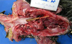
Spotty liver syndrome
Spotty Liver syndrome was suspected in a free-range layer flock of 12,000 hens aged 28 weeks. Sporadic losses and an approximate 6 per cent reduction in egg production were reported over the previous week. Gross pathological examination of three affected hens found similar lesions - perihepatitis and multifocal 1-2mm white lesions, confirmed as a miliary necrotic hepatitis by histopathology. Microbiology investigations were unrewarding as has been the experience since the same putative syndrome was recognised some 50 years ago. Although Campylobacter species infection has been suggested as the cause of so-called ‘avian vibrionic hepatitis’, the aetiology remains elusive.
Broilers and Broiler Breeders
Yolk sac infection (omphalitis)
Increased mortality in a group of 21,800 broiler breeder chicks which had been on the premises for 48 hours was attributed to yolk sac infection associated with E. coli infection. Post-mortem findings included congested yolk sacs and variable amounts of fluid and fibrin in the abdominal cavity with fibrinous pericarditis, splenic enlargement and excess urates in the ureters.
Gastrocnemius tendon rupture
Five 28-week-old broiler breeders in a group of 87 became lame. Treatment was unrewarding and one bird was submitted for post-mortem examination. Necropsy revealed a partial unilateral rupture of the gastrocnemius with associated haemorrhage and inflammation of the muscle fascia. Rupture of the gastrocnemius tendon is recognised in broiler breeders and the aetiology may be attributable to biomechanical stress.
Turkeys
Respiratory aspergillosis (Brooder pneumonia)
Two laboratories diagnosed respiratory aspergillosis in young purchased turkey poults. In one case, losses of 30 poults a day over the first week were reported from 3,400 birds. Characteristic post-mortem findings comprising a multifocal fungal pneumonia and airsacculitis were identified and Aspergillus fumigatus was isolated from the lesions.
Cardiohepatic syndrome of turkey poults
Sudden death of 15 of 42 turkey poults over a 10 day period prompted investigations. Three-week-old casualty birds were examined post-mortem and showed excess serous fluid in the pericardial sac and abdominal cavity with pulmonary congestion and oedema. Histopathology confirmed features consistent with cardiohepatic syndrome. Typically, incidence peaks at two weeks of age and disappears at three weeks. Rapid early growth and environmental conditions are thought to be predisposing factors.
Oesophageal perforation
A five-week-old turkey from a housed group of 300 was found dead. Two other turkeys from the group had died and on-farm post mortem revealed metal staples within the gizzard. Necropsy of the submitted turkey revealed a fibrinous polyserositis and a 3mm hole in the caudal oesophagus, through which a 10cm length of straw protruded. It was concluded that the turkey died as a result of the sequelae to the oesophageal perforation which might have been caused by the ingestion of metal staples.
Backyard Flocks
Infectious laryngotracheitis
Post-mortem examination of a Silkie bantam that had been found dead showed airsacculitis, pleurisy and mucus and blood clots within the trachea. In combination with histopathology, the findings were considered suggestive of Infectious Laryngotracheitis.
Red mite
Severe red mite (Dermanyssus gallinae) infestation was diagnosed in a small backyard flock of former commercial layer hens. Post-mortem examination of one casualty revealed conspicuous carcase pallor consistent with profound anaemia. Three weeks previously, five hens were added to the flock and subsequently, six birds died. It was speculated that the red mite infestation may have been introduced with the new additions.
Gamebirds
Erysipelas
Erysipelas infection was diagnosed following investigation of 50 deaths from a group of 300 eight-week-old pheasants. Pure Erysipelothrix rhusiopathiae growths were isolated in septicaemic distribution. Acute outbreaks of erysipelas are not uncommon in climatic housing and free-range conditions and may involve a history of indirect contact with pigs or sheep. The organism is ubiquitous in the environment and factors such as feather loss or skin damage may predispose to an increased risk of infection.
Suspected Heterakis isolonche typhlitis
single 8-week-old pheasant poult was received from a site where 22 of 400 birds reportedly died and most were showing poor growth. Post-mortem examination findings included pale necrotic material in the caeca accompanied by watery large intestine contents with necrotic debris. Wet smear examination of caecal contents revealed profuse numbers of motile trichomonad protozoa and immature HeterakisEnteric protozoal disease spp. worm larvae. It was considered that the latter were Heterakis isolonche, which is severely pathogenic in pheasants (unlike the more common Heterakis gallinarum which is not usually considered pathogenic). Unfortunately, no adult Heterakis worms were present to confirm the identification, but H. isolonche is a recognised cause of caecal necrosis and fatal nodular typhlitis. Hence, a provisional diagnosis of H. isolonche infection was made, a condition rarely seen in pheasants in recent years.
Enteric protozoal disease
Enteric protozoal disease was commonly diagnosed in 6- to 14-week-old pheasant and red-legged partridge poults. Presenting signs included poor body condition, dehydration and mortality. In the younger poults diagnoses of coccidiosis were common. At post-mortem examination liquid haemorrhagic small intestine contents and caecal cores were reported. Co-infections of coccidiosis with motile protozoa (trichmonad and Spironucleus spp.) were also identified in 6-10 week old poults. In one flock of 20,000 pheasants aged 6-weeks the loss of 4,000 birds was attributed to coccidiosis and spironucleosis (formerly hexamitosis). In older birds, aged 8-14 weeks and often housed in release pens, spironucleosis was commonly diagnosed. At post-mortem liquid small intestine contents and abnormally distended caeca were seen, sometimes with characteristic yellow frothy contents. In one case, ten per cent of a group of 5,000 11-week-old pheasants showed weight loss and abnormal faeces and spironucleosis was diagnosed. Older game birds are less likely to show evidence of coccidial disease due to the common practice of in-feed anticoccidial medication. Therefore, Spironucleus is more commonly detected as an enteric pathogen in this age group. There is currently no licensed medication available for motile protozoa infections in game birds and control hinges on attention to stocking densities and maintaining good biosecurity and hygiene standards.
Further Reading
| - | You can view the full report by clicking here. |
| - | Find out more information on the diseases mentioned in this report by clicking here. |