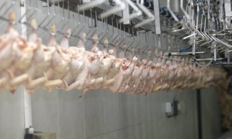
AHVLA: Wet Litter in One of Six Broiler Houses
UK - Wet litter was reported in one of six houses of 42,000 broilers aged 25-days. Deterioration in litter quality was first noticed around 23 days of age, when there had been a slight drop in feed and water consumption which had then returned to normal, reports the latest AHVLA Scanning Surveillance Report dated July 2012. 9 October 2012
 4 minute read
 By:
By: Two sheds had been similarly affected in the
previous crop. At postmortem examination of eight birds, brown watery faecal staining of the vent was
evident with small intestinal and caecal distention with excess fluid content.
Histopathology of
intestine showed moderate chronic active enteritis suggestive of an antigenic response to luminal
antigens. Evidence of Infectious Bronchitis virus (IBV) was also detected from four birds by RT-PCR
testing of tracheal swabs and an IBV variant with 89.3 per cent similarity to 4/91 (793/B) was identified.
It is
often not possible to definitively establish the cause of episodes of wet litter and the cause may
frequently be multi-factorial.
Turkeys
Starve out
Several submissions of 4- to 7-day-old turkey poults were received from small-scale seasonal
producers (100-450 birds) to investigate excessive losses of up to 20 per cent. Consistent findings at
postmortem examination included an absence of feed in the crop and proventriculus and the
presence of bedding material in the gizzard.
Intestinal tract contents were sparse with distended gall
bladders. These findings are typical of so-called starve-out. This is typically caused by a flock
management failure leading to birds not feeding during the first days of life following placement.
Mortality ensues as yolk sac reserves are depleted around 4-days-old. Appropriate advice was
provided.
Marek’s disease
An adult turkey stag was submitted from a smallholding comprising a large number of different
species of birds. The turkey stag had been found dead after an episode of acute enteritis that was
apparently unresponsive to antibiotic treatment. At postmortem examination widespread systemic
visceral abscessation and mycosis were observed as well as marked liver and kidney enlargement
with a mottled appearance.
Histopathology identified pleomorphic lymphocyte infiltration typical of
Marek’s disease, which was subsequently confirmed by PCR. It was proposed that infection with Marek’s disease virus, possibly transmitted from other gallinaceous poultry on the premises, had
debilitated the stag to allow opportunistic infection with a variety of other pathogens leading to its
demise.
Ducks and Geese
Erysipelas
Approximately 200 Mallard ducks aged six weeks from a flock of 5,000 had died over a four day period. Younger birds and adult parent stock were reported to be unaffected. Most birds were found dead and some presented with signs of malaise and bloody nasal discharge. Postmortem examination findings were indicative of septicaemia with Erysipelothrix rhusiopathiae identified by culture, consistent with a diagnosis of acute erysipelas.
Goose parvovirus
Day old goslings had been purchased from a breeder farm. Losses
started at 14 days of age, preceded by acute onset of clinical signs
with rapid deterioration over a 24-hour period. Signs included
inappetance with a reluctance to drink, huddling, vocalising, having
difficulty rising from a sitting position and shaking their heads.
Sixty
of the 117 goslings died over a 2-week period. A batch of 107
goslings purchased five weeks previously from the same supplier
presented similarly, and approximately 70 had died. Another batch
from a different source and housed separately was not affected by
8 days of age.
No illness was reported in other species of poultry
on the farm. Live goslings were submitted for investigation and they
were all showing characteristic clinical signs of goose parvovirus
(GPV) infection including weakness and reluctance to stand,
mucoid brown oculonasal discharges with periorbital crusting and
intermittent head-shaking resulting in feather staining.
Following postmortem examination a diagnosis of GPV was confirmed by identification of typical
histological findings and virus isolation (Irvine and Holmes, 2010). GPV outbreaks have been
diagnosed periodically in Great Britain (GB) since 2004 (VIDA, 2011).
For further information about GPV see the VLA website.
Backyard flocks
Fowl cholera
Respiratory disease and malaise were reported affecting one of four groups of 50 free-range layer
hens. Approximately 40 hens were initially affected with mucoid and catarrhal conjunctivitis, swollen
infra-orbital sinuses and prostration. The owner had elected to cull the affected birds and the issue
appeared to resolve.
However, two weeks later disease recrudesced in the remaining hens from this
group prompting the submission of an affected bird for investigation. Postmortem examination
findings comprised excess serous fluid in the upper respiratory tract, catarrhal material in the sinuses
and fibrinous airsacculitis and pericarditis. Pasteurella multocida was isolated, confirming a diagnosis
of Fowl cholera.
Game birds
Enteric diseases
With the game bird rearing season well underway, numerous submissions of pheasant and redlegged partridge chicks and young poults were received for investigation of increased mortality and/or
enteric disorders. In one case rotavirus infection was confirmed in five-day-old pheasants reported as
showing unevenness with 10 per cent mortality. Spironucleosis (Hexamitosis) was most commonly
diagnosed affecting poults aged 7-10 weeks, with concurrent infections including Syngamus trachea
(gapeworm) in several flocks and Blackhead in a further case.
Husbandry problems were also
identified as contributory factors. Low brooding temperatures and excess humidity were identified as
the cause of approximately 90 per cent of 2000 pheasant chicks aged 14-days to be huddling under heaters
with concurrent rotavirus infection detected. In a separate case, feeder and drinker positioning relative Page 7 of 7
to heat sources led to the loss of at least 600 of 2000 red-legged partridge chicks by 5-days of age
due to starve out.
Salmonella Pullorum
Increased mortality in 11-day-old pheasant chicks resulted in the detection of Salmonella Pullorum. Abnormal faeces and ‘cheesy’ caecal cores were described. Disease caused by S Pullorum is now rare in GB, with only a very small number of isolates obtained each year, typically from game bird or backyard flocks. The organism is host-adapted and hence not considered to be of public health significance. Vertical transmission has an important role in the perpetuation of infection.
Further ReadingFind out more on the diseases mentioned in this report by clicking here. |