


AHVLA: Erysipelas Hits Free-Range Egg Flock
UK - An increase in mortality, of up to 90 birds per day, was occurring in 60-week-old free range layers, with up to 3,000 birds having died in total since placement in a house of 16,000, according to AHVLA's Disease Surveillance Report for September 2012. 12 December 2012
 5 minute read
Commercial Layers
Erysipelas
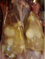
An increase in mortality, of up to 90 birds per day, was occurring in 60-week-old free range layers, with up to 3,000 birds having died in total since placement in a house of 16,000. One other house was relatively unaffected. Postmortem examination showed that the birds had enlarged spleens, mottled kidneys, they were in relatively poor condition and several had a marked peritonitis with yellow cloudy fluid in the peritoneal cavity. Erysipelothrix rhusiopathiae was consistently identified and histopathology revealed chiefly coagulative necrosis of the spleens and livers consistent with bacterial septicaemia.
Spotty liver syndrome
The carcases of two 32-week-old laying hens were submitted as part of an investigation into grumbling mortality with 20 deaths from approximately 5,000 free range hens. Postmortem examination showed pale mottled livers in both birds with miliary pinhead size pale lesions throughout the liver substance. The spleen in both birds was not enlarged but had a mottled appearance on the cut surface. Both birds were actively in lay. Intestinal smears showed the presence of Heterakis and Capillaria species worms. Liver tissue cultures were unrewarding. Histopathological examination confirmed a necrotising hepatitis and diffuse splenitis typical of spotty liver syndrome but Gram-stained sections revealed no organisms associated with the necrotic lesions. There is continuing interest in this disease for which there is no clear aetiology and the AHVLA would be interested to hear of any cases seen or diagnosed in practice (Crawshaw & Irvine 2012). Further information, including a case series study questionnaire that is available for owners of affected flocks or their vets to complete, can be found at: http://vla.defra.gov.uk/science/sci_spotty_liver.htm.
Broilers & Broiler Breeders
Adenoviral ventriculitis
Thirteen-day-old commercial broilers showed an increased mortality, with 100 recorded deaths in a house of 45,000 birds. A practitioner postmortem examination showed pale carcases with dark liquid material in the crop and proximal small intestine, severe damage to the lining of the gizzard and occasional hepatitis. Adenoviral infection of the gizzard was diagnosed by histopathological examination and virus isolation. Histopathology revealed a severe acute granulocytic ventriculitis with glandular necrosis and intranuclear inclusion bodies and acute focal necrosis in the liver. The liver lesions were assumed to be secondary to the severe ulceration in the gizzard and bacterial invasion into the portal circulation. Virus isolation was carried out from pooled viscera from the affected birds and identified an adenovirus.
Encephalomalacia in broiler chicks
Enterococcus hirae septicaemia and encephalomalacia in combination with E. coli septicaemia was seen in a flock of five-day-old broiler chicks submitted with a history of slightly increased mortality. The postmortem findings included swollen livers and spleens from which E. coli and E. hirae were cultured. Histopathological examination of the brain in one bird from which Enterococcus hirae was isolated revealed lesions of encephalomalacia in the brainstem, consistent with E. hirae associated encephalomalacia. This is a self limiting condition which tends to occur in chicks between three and eight days of age, it is associated with transient mortality and neurological signs although these were not reported in this submission.
Turkeys
Marek’s disease
An initial investigation at 49 days of age followed reports of 12 turkey poults in sternal recumbency. Subsequent postmortem examination of casualty birds revealed enlarged livers and spleens with conspicuous focal lesions. Over the following four weeks, 90 out of 600 poults died. Histopathological examination revealed pleomorphic lymphomatous lesions suspicious of Marek’s disease, and this diagnosis was subsequently confirmed by PCR testing. It is unusual to diagnose Marek’s disease in turkeys; the most likely source of infection was some chickens kept on the same holding. Outbreaks of Marek’s disease in turkeys are occasionally reported when the birds have direct or indirect contact with chickens on the same premises. This also represents the second case of Marek’s disease in turkeys investigated in different poultry flocks by AHVLA in the last six months.
Blackhead
Blackhead was diagnosed by Luddington in 11-week-old turkeys. Ten of 230 birds at risk died over a one week period, and several birds had yellow caecal droppings. Postmortem examination of three casualty birds revealed extensive hepatic focal necrotic lesions and necrotic typhlitis characteristic of Histomonas meleagridis infection. Interestingly, following difficulties experienced last year on a free range system, this year birds had been housed in a barn with a concrete floor. However, no worming regime had been adopted as part of preventative measures to control Heterakis sp worms that can harbour the H. meleagridis parasite.
In a separate incident investigated by Winchester, two organic, five-month-old turkeys were submitted following the death of eight turkeys out of a flock of 375. Postmortem examination of one of the birds revealed numerous 1-2 cm diameter pale nodular lesions throughout the liver tissue and one of the branches of the caecum contained a necrotic core, and the other branch contained yellowish fluid content. In the other bird, the liver was unremarkable but there were marked adhesions between the intestinal loops and a necrotic peritonitis. Histopathological examination of the liver lesions from the first bird revealed a marked chronic granulomatous hepatitis with fibrosis, and protozoa were visible consistent with Histomonas sp (blackhead). The peritonitis in the second bird may have resulted from perforation of the caecum associated with blackhead. The liver lesions were almost tumour-like in appearance, which can be a feature of the chronic stages of blackhead in turkeys.
Further ReadingFind out more information on the diseases mentioned here by clicking here. |