

Normal Birds - A Review of Avian Anatomy
By F. Dustan Clark, Extension Poultry Veterinarian, Center of Excellence for Poultry Science at the University of Arkansas's Avian Advice - A necropsy is the examination of a bird externally and internally to determine the cause of death. The method for doing a necropsy varies and depends somewhat on the bird involved, the preference of the individual performing the necropsy, the disease(s) suspected, and where the necropsy is being done.| The Author | |
 Dr. Dustan Clark Extension Poultry Health Veterinarian |
|
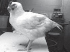
Regardless of the method; the most important point to remember is to systematically evaluate each organ and organ system for changes associated with disease. Since only a few diseases cause very specific lesions in the organs; it is very important to be “familiar” with the normal external and internal anatomy. Usually a necropsy starts with a detailed examination of the external anatomy of the bird.
External Anatomy
Feathers and Skin
Feathers cover the majority of the skin
and are arranged in feather tracts rather than
randomly distributed. The feathers should be
clean at the point of attachment to the skin
and the edges of the feathers should be
smooth with no clear areas present in the
barbs.
The skin of a chicken and/or turkey is
thin and semi-transparent over most of the
body. The muscles, veins, and fat deposits can
be observed through the skin in most birds.
The muscles appear as dark areas; whereas,
fat is yellow. The skin on the face and bottom
of the foot is thickened and is normally white
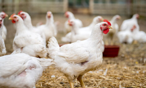
or yellow in color. The comb, wattles, and
car lobes are usually bright red in color in
commercial layers and broiler breeders. It is
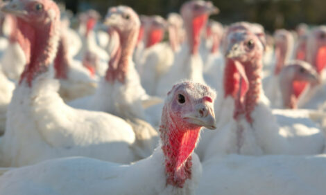
normal for market and breeder turkeys to
develop red or bluish skin on the head and
neck. Normal commercial layers and breeder
hens may have a reddish yellow skin on the
comb, ear lobe, or other facial structures (this
is especially true if they are beginning to
come into production or are out of
production).
 The lower legs are covered with scales
which are yellow to white in coloration. The
thickened skin on the bottom of the foot
(footpad) is usually a pale yellow-tan or
yellow-white color (the scales of the leg arc
similarly colored). Chicks and poults have
yellow colored leg scales. Adult broilers and
commercial layers can have yellow or white
leg scales. Turkey leg scales are white to light
tan colored. The leg coloration will change in
hens from yellow to white and vice versa as
they go into or out of egg production.
The skin, leg, and feather coloration of
many of the varieties of chickens, ducks, and
turkeys kept as backyard, hobby, pet, or
exhibition flocks may vary from those listed.
The best source for individual breed
differences is the book “ The American
Standard of Perfection,” which is published
by the American Poultry Association or the
“American Bantam Standard.”
Ears, Eyes, Nostrils and Beak
The ear in a bird is covered with fine
feathers and is a small opening located on the
side of the head. The eye should be a bright
yellow-orange in color and free of discharges. The eyes should
be clear with dark black pupils surrounded by a colored iris.
The color of the iris varies with the breed and age of the bird,
but in general is steel-grey in chicks and poults. In adult
broilers, layers, and broiler breeders the iris is yellow-orange;
but brown in adult turkeys. The nostrils are slit like openings
on top of the beak and at the base of the beak. They are
surrounded by tan-yellow fleshy skin called the cere. The beak
is a yellow-horn to white-horn color in the normal bird and has
a smooth surface with the end of the beak pointed or blunted
in a beak-trimmed bird. Again, colors other than those listed
may be normal for many of the varieties of chickens, ducks,
and turkeys kept as backyard, hobby, pet, or exhibition flocks.
As before, the best source for these breed differences is the “
The American Standard of Perfection” or the “American
Bantam Standard” book.
Internal Anatomy
Once the external anatomy has been evaluated the
internal anatomy of the bird is examined. The skin should be
removed and the bird opened to expose internal organs. The
procedure of initially opening the bird to evaluate the internal
organs may vary depending on the personal preference of the
individual performing the necropsy. However, regardless of
the procedure, it is important to evaluate all organs present
systematically and thoroughly.
The first organs that come into view when the skin of a
chicken or turkey is removed for necropsy are the muscles,
sternal bursa, and bone (keel). The breast muscles are a greywhite
in color normal poultry. The point of the keel is white
and the edge of the bone is straight. The sternal bursa is a
white sac-like structure that is located on the sternum and
contains a small amount of clear fluid. If the leg muscles are
observed they are a darker grey-white color and the sciatic
nerve (located between the leg muscles) is a glistening white
with cross striations.

Thoracic (Chest) and Abdominal Anatomy
After the sternum and breast muscles are removed the
internal organs are evaluated, The heart is a triangular shaped
organ (the base of which is toward the head of the bird) that is
surrounded by a clear sac (pericardial sac). The heart is greywhite
in color and has a band of yellow fat near the base.
Internally, the heart is the same color with clear membranous
valves between heart chambers. The left ventricle (lower left
chamber) of the heart is thicker than the right ventricle. The
heart is almost completely surrounded by the lobes of the
liver.
The liver is the largest internal organ, is firm, and has
prominent sharply defined edges. The color of the liver varies
with diet. Baby chicks and poults tend to have a liver that is
yellow in color due to yolk absorption. Adult birds can have a
yellow-tan liver if on a high fat diet and the organ may be soft.
The adult bird usually has a dark red to red brown colored
liver.
The avian gallbladder is attached to the liver lobe and can be
easily examined by moving the liver to one side. This sacklike
structure is greenish-black in color due to the bile present
in it.
The trachea and syrinx (voice box) are visible at the base
of the heart. These structures are white with the trachea a
round tube like structure that divides into smaller left and right
bronchi. The syrinx is a flattened area of the trachea that is at
the end of the trachea before it dividing into bronchi. The
bronchi are identical to the trachea in color and shape but are
of a smaller diameter. However, a better examination of the
trachea is done in the neck of the bird. The aorta is also visible
at the base of the heart and is the artery that connects to the
heart’s left ventricle.
This tubular structure is thick walled and
pink white to red-white in color. The aorta and smaller
connecting arteries are better examined after the organs in the
thorax and abdomen are removed. The fat pad that covers the
organs must be cut or torn to reveal the gizzard (ventriculus)
and the stomach (proventriculus) to the bird’s right side. The
spleen is readily visible at the junction of the stomach and
gizzard after they are exposed. This lymphoid organ is oval or
elliptical in shape and dark red to purple in coloration. The
spleen in an adult bird is approximately one inch long.
The air sacs on the left are also readily visible after the
stomach and gizzard are set aside. These clear membranes are
attached to the lungs and increase the respiratory capacity of
the bird. Female birds that are in production may have yellow
fat deposits on the air sacs. The air sacs on the bird’s right side
should also be examined; it is usually necessary to move the
liver, stomach, and gizzard to the bird’s left side to examine
them adequately.
Avian lungs are closely adhered to the ribs and are an
orange-red or pink-red color. The lungs can be removed for a
close examination of the ribs. The ribs, as with all avian
bones, are smooth thin walled and white. Immediately below
the lungs are the kidneys, adrenal glands, and gonadal tissues
(testes or orvaries). The kidneys are firmly embedded in
depressions in the bone (synsacrum) and have three distinct
lobes (cranial, middle, and caudal). The bird has two kidneys,
a left and right, and these organs are dark red to dark brown
with a fine reticulated patient visible. A small, white tube (the
ureter) connects each kidney to the cloaca. The adrenal glands
are small tan triangular shaped glands located at the section of
the kidney near the lung. Gonadal tissue is also located near
the kidney. The male has two testes, one on either side of the
midline.
These organs are bean shaped or elliptical shaped and
tan. Two small white coiled tubules connect the testes to the
cloaca. In the female only the left ovary and oviduct are
generally present near the left kidney. In an immature female
the ovary is roughly triangular in shape or shaped like an
inverted L. It is white to light yellow in color and may have a
granular or gritty appearing surface. The developed oviduct is
a large grey-white tubular organ that has distinct longitudinal
structures. The oviduct connects the ovary to the cloaca and
adds egg components such as albumen, shell membranes and
shells as it transports the follicle (yolk) to the surface.
Located near these organs and near the midline is the
descending aorta. This thick walled artery is a continuation of
the aorta as it leaves the heart. It is from this major artery that
numerous smaller arteries arise to supply blood to the internal
organs. The aorta is pink-white to red-white in color.
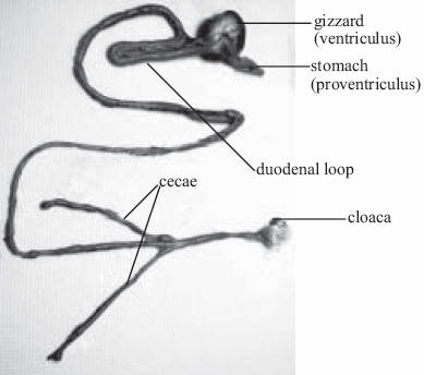
The digestive tract should be examined next. The
stomach (proventriculus) is a spindle shaped organ that has the
gizzard (ventriculus) attached to it. The stomach is grey in
color and internally the lining in glistening grey-white with
small papillae (gland openings) present. The gizzard is a round
dark brown to dark red organ attached to the gizzard.
Internally, the gizzard (ventriculus) has a koilin lining which
is yellow to yellow-green in color.
The duodenum is the first section of the small intestines.
It is loop shaped and surrounds the pancreas. The pancreas is a
white-tan fleshy organ. The duodenum, like all of the small
intestines is a tan-grey to white-grey tube which has a fine
textured lining similar to the surface of a towel. The jejunum
and ilium are the next two sections of the small intestines.
Two sack-like structures are attached to the small intestines at
the junction of the large intestines and ileum. These structures
are the cecae which are thin walled with small thick areas in
the wall (cecal tonsils) at the points of attachment to the small
intestines. Cecal contents are dark green or dark brown. The
large intestine (which is very short) lies between the ileum to
the opening to the surface called the cloaca. The cloaca is
similar in color to the small intestines but is of larger diameter.
Feces in the large intestine and cloaca is generally drier and
green to brown feces in color. The ileum contains a more
liquid feces of similar color. White pasty urates are often
present in the cloaca. The bursa of Fabricius is a round tanwhite
lymphoid organ which is organ located behind (dorsal)
the cloaca.
Most blood vessels are examined along with the organs
such as checking the large vessels coming to or leaving the
heart when the base of the heart and syrinx are examined.
Blood vessels vary in size depending on the organ supplied.
Arteries are thicker walled than veins, and are a pink white to
red white in color. Veins are thin walled, tend to flatten out
when touched and are a dark blue in color due to the blood in
them.
Neck Region
The mouth and neck of the bird should also be examined.
A cut is made at the corner of the mouth and extended down
the neck, thus exposing the structures for closer examination.
In the mouth of the bird the tongue can be examined. This
triangular shaped organ is dull grey-white and has a few
bumps (papillae) on the surface. Directly behind the back of
the tongue (and connected to it) is the glottis. The glottis is the
opening of the trachea. It is white in color and has two folds
(left and right) which come together to close the opening when
the bird swallows. The oropharynx is the region at the back of
the mouth and is a glistening grey-white color. Located on the
roof of the mouth is the cleft opening called the choana. This
structure should be clean with a small amount of clear mucous
usually present in the cleft. The choana is also grey-white in
color and numerous conical papillae are around the cleft.
The esophagus should be opening and examined. It too is
grey-white in color and has a smooth surface. There is an
organ at the base of the esophagus called the crop. The crop is
a pouch of the esophagus and as such is the color and texture
of the esophagus. The trachea is also present in the neck. This
white tubular structure has rings of cartilage visible from the
outside. Inside the trachea is a small amount of clear mucous
and the lining is a glistening clear white.
The remaining most obvious organ in the neck is the
thymus. This organ is multi-lobed and tan in color. Often
yellow fat is intermixed with the lobes. This organ 15 located
near the base of the neck and crop.
The beak should be removed to expose the nasal cavity.
There are scroll like structures in the nasal cavity which are a
tan-white in color. There is also a small amount of clear
mucous on these scrolls.
Another area to examine is the breast musculature. The superficial breast muscle should be cut into to check the deep breast
muscle (supracoracoideus muscle). This deep muscle is the same color as the superficial breast muscle.
The joints of the leg are also cut into and examined. All joints in the leg should contain a clear fluid. The cartilage in the leg
joints can also be examined at this time. Cartilage is a bright white to grey- white in color and has a smooth surface. The ends of
the leg bones are usually cut to examine the bone marrow, and check for cartilage plugs. If a cartilage plug is present in the end of
the tibiotarsal bone it appears as a triangular shaped plug that is white to grey-white in color. Bone marrow is red in color and soft
in texture.
The structures and organs discussed are those examined on a routine field necropsy. Naturally, any area that looks
“abnormal” is more closely examined.
Source: Avian Advice - Winter 2005 - Volume 7, Number 1


