


|
Diseases of Poultry
By Ivan Dinev, DVM, PhD
|
HISTOMONOSIS
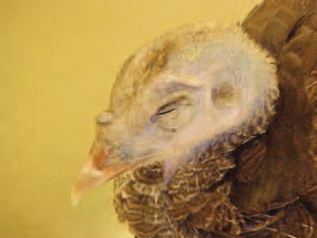
362.. The histomonosis is a protozoan disease, caused by Histomonas meleagridis, and characterized by necrotizing lesions affecting the liver and the caeca. Clinically, sulfur-yellow coloured faeces and depression are observed. A characteristic feature is the blackening of the skin of the head (blackhead), due to cyanosis.
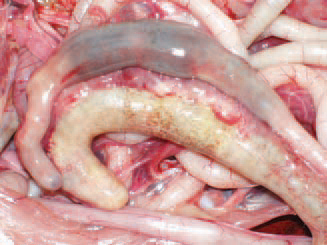
363.Pathoanatomically, bi-lateral enlargement of caeca with thickening of walls is observed. The aetiological agent is Histomonas meleagridis, a polymorphic flagellate that is present as flagellate in caeca and as amoeba in tissues. The trophozoites survive for several hours in the environment but in Heterakis eggs, they remain infective for more than a year.
364.Often, the occurring typhlitis is the cause for adhesive peritonitis.
365.Susceptible species are turkeys, chickens, pheasants, rock partridges, guinea fowl, and geese. The turkeys are the most vulnerable between 3 and 12 weeks of age and chickens between 4 and 6 weeks of age. The caecal mucosa is usually ulcerated.
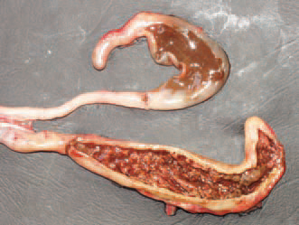
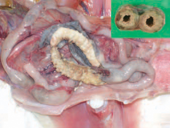
366.. The main vector is Heterakis gallinarum through the eggs, respectively the larvae, where Histomonas meleagridis forms are found. Some wild birds could also serve as vectors. The caecal content is often mixed with blood.
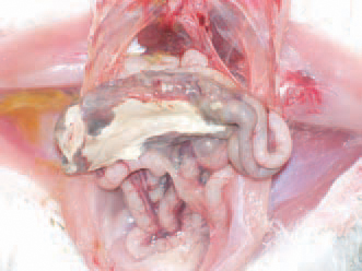
367.In older cases, crusts of dense caseous masses are formed into the carca that thicken this intestinal wall and reduce the lumen (top right: transverse cross section through caeca.)
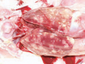
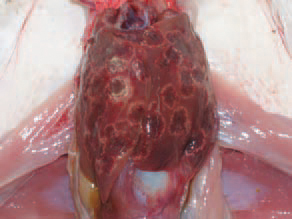
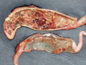
368.369.Earth worms are mechanical vectors of H. gallinarum larvae. The main reservoirs of infection are hens and chickens. The morbidity rate amounts to 90% and the mortality rate to 70%. In the liver, irregularly outlined coagulation necroses with various size and colour, are observed.
370.371.Usually, necroses represent yellowish to grey or red (haemorrhagically infarcted), well delineated oci with diameter of about 1-2 cm. Diagnosis - it is made on the basis of the typical macroscopic lesions. When necessary, a histological study and phase-contrast microscopy of native preparations could be performed. Histomonosis should be differentiated from UE, coccidiosis and alimentary tract trichomonosis (Trichomonas gallinarum), where not counting the caeca, lesions are also present in the last third of small intestine.