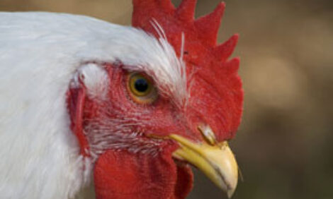
Finding New Methods to Detect Chicken Anaemia Virus
Chicken infectious anaemia has become an emerging threat to the poultry industry worldwide. 2 July 2015
 4 minute read
 By:
By: It is caused by a circovirus called chicken anaemia virus (CAV), and may cause clinical symptoms in young chickens.
Chicken infectious anaemia is a relatively new disease of chickens, first reported in Japan and it is a virus that may cause clinical manifestation only in young chickens free of anti-chicken anaemia virus antibody, but it can maintain subclinical infection in antibody positive commercial chickens and cause immunosuppression.
Vertical transmission of the virus is well documented, and it is also believed to transmit laterally to flockmates, presumably through the faecal-oral route.
Commercial broiler chickens are not usually vaccinated against chicken anaemia virus, but the parent stock are vaccinated to protect their progeny through passive immunity.
In the absence of clinical manifestation, a reliable method for monitoring of chicken anaemia virus infection is needed for the poultry industry.
The detection of the viral nucleic acid in various tissues using molecular techniques is commonly used. However, the virus may not be present in all organs at all stages of infection. In addition, the tissue distribution may depend on the antibody status of the host.
Two experiments on tissue distribution and shedding profiles of chicken anaemia virus in specific pathogen-free and commercial broiler chickens were conducted, one in specific-pathogen-free-layer chickens and the other in commercial broiler chickens, to detect and quantify the viral genome in various tissues and environmental samples.
The researchers M. Alsharari, A.F.M.F. Islam, S.W. Walkden-Brown and K.G. Renz in their paper to the 26th Australian Poultry Science Symposium found that chicken anaemia virus was detectable in a number of tissues with a high titre in thymus and bone marrow and therefore, these two tissues were taken as samples for molecular diagnosis.
The virus was detected in dust and litter samples, although at a low level.
The monitoring of chicken anaemia virus infection using environmental samples such as dust and litter has potential, but needs further optimisation.
The study showed that the chicken anaemia virus genome can be detected in lymphoid organs and bone marrow as early as six days post-infection in maternal-antibody-free chickens but not in commercial broiler chickens.
The authors said that this is the first study to demonstrate that chicken anaemia virus can be detected in dust and litter samples in both pathogen-free and commercial chickens.
This has implications for the poultry industry, as environmental monitoring can be used for disease screening.
“Among the four organs tested, we found that the thymus had the highest viral load,” the research team said.
“The viral copies increased up to 13 days post-infection, following which they either stayed at similar levels (bursa and bone marrow) or increased (thymus and spleen) up to 28 days post-infection in this study, whereas only a limited number of chickens were reported to be chicken anaemia virus positive in the thymus and spleen and none in the bursa and caecal tonsil following infection with a vaccinal virus."
The virus was detectable in the faeces, but at a very low level.
Therefore, the researchers concluded that the shedding of chicken anaemia virus through faeces and the faecal-oral route of lateral infection remain to be determined.
The most significant finding of the study was the detection of the virus in chicken dust and litter.
However, the team said that the viral load in these environmental samples was not very high (103.9–102.6 per mg).
The presence of the chicken anaemia virus genome in various tissues reached its highest level in the bone marrow and the study concluded that bone marrow should be the best sample for diagnostic purposes followed by the thymus and bursa, but not the spleen.
Dust also has potential for use in monitoring the disease, but not litter.
Although this study successfully demonstrated the ability to detect the chicken anaemia virus genome in the dust and litter both from pathogen-free and broiler chickens that had no clinical manifestation of the disease, these methods need further validation before industry recommendations can be made.
The team said that the industry would ideally like one method for the detection of all potential viral diseases from a single sample and this may be best be achieved by applying two different nucleic acid extraction methods, one for DNA viruses and one for RNA viruses.
REFERENCES
Barrios PR, Marín SY, Resende M, Rios RL, Resende JS, Horta RS, Costa MP & Martins NR (2009) Brazilian Journal of Poultry Science 11: 135-138.
Cardona CJ, Oswald WB & Schat K (2000) Journal of General Virology 81: 2067-2075.
Davidson I, Artzi N, Shkoda I, Lublin A, Loeb E & Schat, K (2008) Virus Research 132: 152-159.
Dren CN, Kant A, Van Roozelaar DJ, Hartog L, Noteborn MH & Koch G (2000) Acta veterinaria Hungarica 48: 455-467.
Dye C, Helps CR & Siddell SG (2008) Journal of Feline Medicine and Surgery 10: 167-174.
Islam A & Walkden-Brown SW (2007) Journal of General Virology 88: 2121-8.
Tan J & Tannock GA (2005) Journal of General Virology 86: 1327-1333.
Vaziry A, Silim A, Bleau C, Frenette D & Lamontagne L (2011) Avian Pathology 40: 377- 385.
Yuasa N, Taniguchi T & Yoshida I (1979) Avian Diseases 23: 366-385.
Walkden-Brown SW, Islam A, Groves PJ, Rubite A, Sharpe SM & Burgess SK (2013) Avian Diseases 57: 544-554.
Zhang T, Wang B, Ling Z, Li Y, Gao X, Tian G, Zhang J, Han L & Zhang L (2009) Chinese Journal of Animal Quarantine 26: 58-60.
July 2015