UK Poultry Disease Monthly Surveillance Report (to October 2004)
By Veterinary Laboratories Agency - This report monitors trends in the major endemic poultry diseases and utilises the farmfile and VIDA (Veterinary Investigation Disease Analysis) databases. The report is compiled using disease data gathered by the network of 15 VLA regional laboratories which carry out disease investigation in the field. 21 October 2004
 4 minute read
 Published October 2004 Highlights Blackhead causing losses in free-range layers Fowl pox diagnosed in layer pullets Salmonella virchow causing losses in turkeys |
Salmonellosis
Salmonella Virchow was isolated from eight, four-week-old turkey poults in a
flock where deaths had been encountered over the previous 7 days. In all 519
deaths occurred in two sheds, 137 out of 1200 birds in one, and 382 out of
1700 in another. Just under 10,000 turkeys were kept on this farm in eight
sheds. Other sheds contained birds that were much older and kept for farmgate
sales, and stags for the catering trade.
Birds were being found dead with
little premonitory signs. Necropsy revealed spleen enlargement, airsacculitis,
peritonitis and perihepatitis. White plaques were seen adhered to the
mesentery. The Health Protection Agency (HPA) was informed and an
advisory visit was carried out to advise on the risk of zoonotic infection and
the biosecurity precautions associated with the farm gate sale of older birds.
Blackhead
Histomoniasis (Blackhead) was diagnosed by Luddington on two separate
turkey units following necropsy of affected birds. The units were rearing freerange
birds for the Christmas market with one flock of 500 and the other of
2000 poults (in 4 groups of 500). Clinical signs including malaise, ill-thrift,
yellow diarrhoea and deaths were reported with birds affected at 20 and 14
weeks of age in respective flocks.
Losses of birds at 38 weeks of lay in a free range flock of chickens led to the
submission of several birds to Winchester for post-mortem examination. Two
of the birds submitted showed the presence of white focal lesions in the liver.
In one bird these were 1-3cm in diameter with irregular edges. In this bird a
small area of caecal necrosis was present. In the second bird the liver
showed the presence of white miliary focal lesions and the presence of
necrotic cores in the caecae. Parasitological examination carried out on
intestinal contents from the group of birds submitted showed the presence of
Heterakis, Ascarid and Capillaria worms. Histopathological examination of
tissues showed lesions consistent with histomoniasis.
Interstitial pneumonitis
Post mortem examination of a 9-week-old female duck by Luddington resulted
in the diagnosis of an unusual interstitial pneumonitis. The duck was the
seventh to be lost in five days from one brood. Clinically, malaise was
reported. The other backyard domestic poultry on the premises were
unaffected. Necropsy findings included the duck to be in average condition
with splenomegaly and yellow fibrin deposits over the thoracic air sacs with
caudal pulmonary consolidation seen. No significant microbiology findings
were made to support the suspected differential diagnoses of septicaemia
(Riemeralla, Erysipelothrix or Listeria species infections). Histopathology
identified a subacute, severe interstitial pneumonitis, subacute multifocal
myocarditis and mild periacinar hepatocyte degeneration.
The lesions were consistent with those described by Randall (1987) as an
unusual intracellular infection in ducks. It is suspected that these cases may
relate to a severe protozoal infestation but the organism has never been
characterised (Randall C.J., Lees S., Pepin G.A. and Ross H.M. 1987, Avian
Pathology, 16: 479 – 491.)
Erysipelas
Two turkeys from a unit raising birds for the Christmas market were submitted to RVC for examination following a number of deaths on the unit. The lesions seen were typical of infection with Erysipelas and the diagnosis was confirmed on bacteriological culture of the organism from the viscera. Although the farm had previously had a vaccination policy, the current crop of birds had not been vaccinated.
Vaccine associated lameness
Between 10 and 15% of 6000 laying pullets aged 17 weeks were showing a unilateral lameness, mainly involving the same limb on each bird. This occurred 2 weeks after a vaccine was administered into what was assumed to be the same limb when loaded for transportation. Post mortem examination showed a marked swelling of one limb, with clearly delineated lines of skin discolouration readily visible (Fig. 1). There was an underlying sub-cutaneous oedema, and superficial necrosis of muscle. No other abnormalities were identified. The VMD were informed.
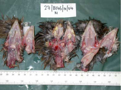
Fowl pox
The loss of 7 of 14 mixed fancy breed, 15-week-old layer pullets over a 2-
week period was investigated. A further 27 free-range layers which mixed by
day with the pullets, but were housed separately overnight, were reportedly
unaffected. Necropsies on three fresh, casualty pullets revealed all to be in
very poor bodily condition (average weight 493g) with multiple, raised, crusted
brown/yellow cutaneous lesions irregularly distributed over the skin of the
head of each pullet. Additionally, irregularly distributed across the
oropharyngeal and rostral oesophageal mucosa of each pullet were multiple,
discrete, pale yellow/cream-coloured, raised necrotic lesions, 2-6 mm in
diameter (see figure 3).
Mucoid “rhinitis and tracheitis” were also seen. The
morphological diagnosis of fowlpox (cutaneous “dry pox” and oropharyngeal
fibrinonecrotic “wet pox” forms) was endorsed by histopathological
examinations which revealed severe, ulcerative dermatitis with granulomatous
inflammation and intracytoplasmic inclusions suggestive of pox virus.
Recommendations included culling severely affected birds, close monitoring
of the remaining pullets for the presence of typical pox lesions, attention to
hygiene and flock anthelmintic treatment.
To read the full report please click here (PDF)
Source: Veterinary Laboratories Agency - October 2004