

UK Poultry Disease Quarterly Surveillance Report - July- September 2008
Highlights of the latest quarterly report from the Veterinary Laboratories Agency (VLA) include fungal infections in young game birds, acute respiratory disease of unknown aetiology in ducks, acute and atypical ILT outbreaks in smaller flocks and the third isolation of QX strain of IBV from a backyard flock.
 July – September 2008 Published November 2008 Contents INTRODUCTION TO GB REPORT OVERVIEW - FACTORS INFLUENCING DISEASE AND SUBMISSION RATES - NOTIFIABLE DISEASES - FARM VISIT INVESTIGATIONS - FOOD SAFETY INCIDENTS - ZOONOSES ENDEMIC DISEASE SURVEILLANCE MISCELLANEOUS AVIAN DISEASE TOPICS |
Highlights
- Wet summer weather adversely affects young game birds and leads to increased fungal infections - Damp feed and bedding led to outbreaks of aspergillosis in poultry and game birds.
- Acute respiratory disease of unknown aetiology in ducks – A previously described disease associated with basophilic intracellular bodies.
- Acute ILT outbreak prompts suspect notifiable disease report - ILT can be controlled by vaccination in commercial flocks. ILT is becoming more common in smaller flocks and may not be recognised by practitioners when the clinical presentation is atypical.
- Third isolation of QX strain of IBV from a “backyard” flock - These episodes highlight the presence of this IBV variant within the poultry population. Extent of virus circulation is unquantified; hence the threat posed to the commercial poultry sector is unknown.
- Increased cases of staphylococcal tenosynovitis in pheasant poults – Some outbreaks appear to be associated with removal of plastic ‘bits’ prior to entry to release pens.
Economics of the Poultry Industry
- Placings
chick placings (average monthly figures)
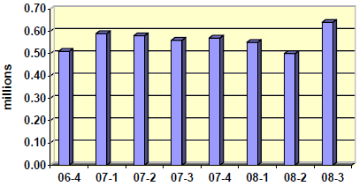
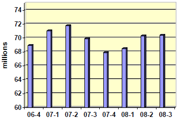
UK quarterly figures for commercial layer chick
placings (average monthly figures)
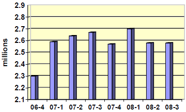
UK quarterly figures for commercial broiler chick
placings (average monthly figures)
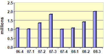
UK quarterly figures for turkey poults
placings (average monthly figures)
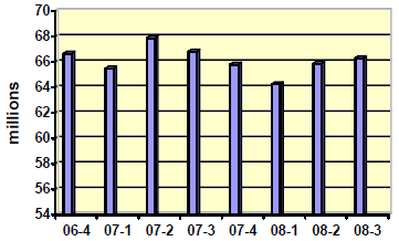
- Slaughterings
slaughterings (average monthly figures)
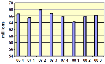
UK quarterly figures for broiler
slaughterings (average monthly figures)
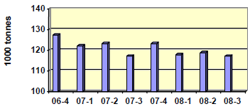
- Meat production
(Average monthly figures)
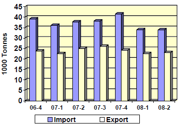
Total Poultry Meat Trade
(Average Monthly figures)
Zoonoses
Salmonella
| In the tables and figures below, an incident is defined as ‘the first isolation and all subsequent isolations of the same serovar or serovar and phage/definitive type combination of a particular Salmonella from an animal, group of animals or their environment on a single premises, within a defined time period (usually 30 days). |
No clinical cases of disease due to S. Enteritidis have been recorded on VIDA in chickens during the quarter, or since 2004 when the last case was recorded.
Sampling of chicken layer flocks according to the requirements of the Salmonella National Control Programme (NCP) for layers is ongoing. More details on the Salmonella NCP in layers can be found on Defra’s web site.
| Table 5. The annual incidents of S. Enteritidis and S. Typhimurium in turkeys | |||||
| 2004 | 2005 | 2006 | 2007 | 2008 (Q1, 2 & 3) |
|
|---|---|---|---|---|---|
| S. Enteritidis (total) |
0 | 0 | 0 | 0 | 0 |
| S. Typhimurium (total) |
38 | 23 | 38 | 12 | 1 |
Note: The incidents of S. Enteritidis and S. Typhimurium exclude isolates arising from the 2006/07 EU survey of turkey flocks (see Avian Quarterly Report, Vol. 10, No 3, July-September 2006, Appendix 1).
| Table 6. The annual incidents of S. Binza and S.Orion in pheasants | |||||
| 2004 | 2005 | 2006 | 2007 | 2008 (Q1, 2 & 3) |
|
|---|---|---|---|---|---|
| S. Binza (total) |
4 | 10 | 21 | 7 | 6 |
| S. Orion (total) |
1 | 3 | 3 | 2 | 2 |
Endemic Disease Surveillance
Commercial Layers and Layer Breeders
Red mite
Several cases of severe anaemia resulting in death caused by red mite (Dermanyssus gallinae) infestation were seen in both backyard and commercial free-range layer flocks.
The red mite is the most significant parasite of layer chickens in the UK, with effects on bird welfare and production. Control is difficult and is currently aimed at reducing the numbers of mites in the birds’ environment. The mites are prolific, being able to complete their life cycle in as little as seven days, acaricides do not penetrate easily into its hiding places and some strains have developed resistance to some acaricides. (see “Poultry Mites: Current European Research” by Peter Bates., Avian Quarterly Report Vol. 5 No 4, October–December 2001) and “The Red Mite Problem”, Dr Bhushy Thind (CSL, York), Avian Quarterly Report Vol. 9, No 2, April-June 2005). Recent research has been aimed at development of a vaccine (a paper entitled “Poultry red mites – a view to vaccination” is to be presented at the BVPA Winter meeting 2008 by workers from the Moredun Research Institute).
Broilers and Broiler Breeders
Wet litter problems
A number of submissions of live birds from flocks aged between 20 and 30 days experiencing wet litter problems were examined during this quarter. In addition, several batches of fixed tissues for histopathology were received from poultry veterinarians investigating this type of problem. In some cases birds showed chronic bursal atrophy which, though not specific, would be consistent with a previous Infectious Bursal Disease (IBD) challenge (birds often scour for a few days and the litter deteriorates during episodes of acute IBD), but in one case birds from a different house on the same site also experiencing litter problems showed no evidence of bursal atrophy or possible IBD. The intestines showed no consistent histological abnormalities (possible viral or bacterial enteritis was not detected). Coccidial infection was present to varying degrees in some cases, but again was inconsistent and in some cases there was clearly little or no coccidial involvement. A reovirus was also isolated from the intestinal contents of 28-day-old broilers with severe diarrhoea, where other infectious and nutritional causes of enteric disease had been excluded.
“Spiking mortality”
This condition has been seen only sporadically since the early 2000s. However, during this quarter postmortem submissions and formalin fixed tissues for histopathology have been received from several episodes where sudden sharp increases in mortality have occurred in broiler flocks usually of around 20- 30 days of age, which had been doing well and had no history of disease or mortality. Findings at postmortem were of birds dying in good condition, often with little or no feed in the upper digestive tract. In contrast to most cases of “spiking mortality” seen in the UK in the early 2000s, some birds in the current episodes have had small amounts of feed in their crop. Many birds showed striking pink colouration of the subcutaneous fat, which intensifies on exposure to air, slight yellowish discolouration of the heart muscle in contracted hearts with prominently congested surface blood vessels, congested livers and less often, congested lungs, and variable pale areas in liver and kidney. Some birds had pale slightly shrunken spleens with patchy “blood-splash” haemorrhages. Conventional histopathology has shown no consistent abnormality. Fat stains demonstrate abnormal quantities of fine lipid droplets in heart muscle and in tubular epithelium of kidney - consistent with mobilisation of fat, as is seen in birds with classical “Fatty liver and kidney syndrome” (FLKS). Significant amelioration of “spiking mortality” episodes in the early 2000s was achieved by increasing the length of the dark period - presumably allowing birds to adapt to periods of fasting, so that they would not become hypoglycaemic if there was accidental interruption to the feed supply. The cause of the recent increased incidence of these episodes in the UK is not known.
Staphylococcal arthritis
Staphylococcal infection caused lameness and increased mortality in two placements of broiler breeder cockerels aged 8 and 13 days. In some affected chicks, histological examination of the toe trimmed at day-old in the hatchery (the tip of digit 1, the “thumb”) revealed foci of coccoid bacteria consistent with staphylococci beneath the healing wound.
Ulcerative enteritis
Increased mortality in a house of 4,000 25-week-old broiler breeder hens was found to be due to Ulcerative enteritis. Gross and histological lesions were consistent with this condition (also known as “Quail disease”). However, attempts to isolate the causative organism (Clostridium colinum) proved unrewarding. Mortality stopped abruptly and did not recur when the flock was given a course of broad-spectrum antibiotic (amoxicillin). This condition is very unusual in chickens of this age.
Turkeys
Aspergillosis
An increased number of cases of aspergillosis were seen in turkeys (as well as pheasants, ducks and geese) this quarter suggesting a particularly heavy challenge with fungal spores. This may relate to the use of poor quality bedding, which will have been exacerbated by the humid conditions experienced this summer. On one farm, fine head tremor was described clinically in 9- to 10-week-old turkey poults. Fungal pneumonia and airsacculitis, mycotic encephalitis, retinitis and iriditis were also present. Mycotic pneumonia due to Aspergillus spp. also caused a cumulative mortality of 20% in a batch of 2000 14-day-old free-range turkeys and the increased mortality seen in a batch of turkey poults from two days of age. Postmortem examination revealed multiple small pale nodules in the lungs of some birds, while some birds had a moderate airsacculitis.
Producers will need to be particularly vigilant for mould contamination of bedding during the coming months if further cases are to be avoided.
Airsacculitis/colisepticaemia
Airsacculitis associated with a colisepticaemia occurred on a number of occasions throughout the quarter. On one multi-age site of commercial meat turkeys, unevenness, stunting and increased mortality were seen from four weeks of age onwards in several batches of birds. Colisepticaemia was a common post mortem finding but some birds had a caseous infraorbital sinusitis and airsacculitis. An unvalidated RRT-PCR test for avian metapneumovirus (aMPV) was carried out on a number of oropharyngeal swabs and one swab from 5-week-old birds (that had not been vaccinated for aMPV) was positive. On another farm, swollen heads were described in 10- to 14-week-old poults. Birds had distension of the infraorbital sinuses, which contained frothy clear mucus, and isolates of Escherichia coli and Pseudomonas aeruginosa were made from various sites. Possible initiating agents in such cases include aMPV (turkey rhinotracheitis virus), Mycoplasma spp., and Ornithobacterium rhinotracheale.
Coccidiosis
Clinical coccidiosis leading to the formation of caecal casts was seen on a number of occasions. One outbreak in 11-week-old turkeys appeared to have precipitated clostridial necrotic enteritis. Caecal coccidiosis, seen in 6-week-old poults, was associated with a return to starter feed when their current feed ran out.
It is rare to see severe clinical coccidiosis causing caecal casts in commercial turkeys on coccidiostats. The humid conditions experienced this year may have led to ideal conditions for coccidial sporulation.
Avian tuberculosis
Avian tuberculosis was responsible for ill-thrift and sudden death on a site with several different breeds of turkey. Miliary lesions were seen on post-mortem examination in the liver, spleen and abdominal connective tissues of two affected birds. Ziehl Neelson-stained smears revealed acid-fast bacilli typical of Mycobacteria and consistent with a diagnosis of avian tuberculosis.
Ducks and Geese
Aspergillosis
Heavy losses in eight- to nine-day-old ducklings were shown to be due to aspergillosis. Although early cases displayed only reddening of lung tissue, and only one bird had miliary lung lesions, Aspergillus fumigatus was isolated from all lungs cultured. Further dead birds examined on-farm showed the typical gross lesions of mycotic pneumonia. Poor quality bedding had been used, and it was considered likely that heavy fungal spore contamination of the environment had led to the incident.
Riemerella anatipestifer infection
Riemerella anatipestifer infection was the cause of death or culling in 30 out of a group of 1,350 5-week-old goslings over a three-day period. They had been out at grass for seven days and presented as collapsed birds, unable to walk with watery diarrhoea and a slight head tremor. Lesions demonstrated included extensive pericarditis, perihepatitis and airsacculitis and Riemerella anatipestifer was isolated from lungs and airsacs. Riemerella anatipestifer infection takes place through the respiratory tract or through wounds on the skin, particularly the feet. Predisposing factors include stress, such as overcrowding, inadequate ventilation, poor hygiene and exposure to adverse weather.
Acute respiratory disease of unknown aetiology
A sub-acute diffuse pneumonia associated with basophilic forms was diagnosed in a domestic duck from a small backyard flock. Such forms have been seen previously in ducklings as an overwhelming infection of lung tissue and this organism has never been accurately identified. In this case there had been one adult duck and one duckling fatality from a group of around 25 ducks on the holding. The submitted duck had been ill for a few days prior to death with a non-specific malaise and loss of appetite. The condition was first reported in the UK in 1987 and, in the intervening years, the VLA and SAC have seen a number of cases of this apparently overwhelming infection, usually in ducklings.
The condition was first described in Canada in 1980 (Julian and Galt, 1980. Journal of Wildlife. Diseases, 16, 39-44) and first reported in the UK in 1987 (Randall et al., 1987. Avian Pathology, 16: 479-491). The organism is thought to be a yeast-like eukaryote. It is apparently widespread in North America, Europe and New Zealand, and occurs primarily in Muscovy ducks, but can also affect other species.
Duck Viral Enteritis (duck plague)
Duck Viral Enteritis (DVE) was the cause of a sudden rise in mortality in 5 to 6-week-old ducklings on a commercial unit. Characteristic diptheritic and necrotic intestinal changes were seen at postmortem examination and large numbers of coccidia were demonstrated on wet smears. However, a herpesvirus typical of DVE virus was subsequently isolated from tissues. There was no known contact with wild birds and the source of the virus could not be established.
Duck Viral Enteritis remains a seasonal disease of waterfowl, with episodes most commonly occurring during the spring months through to June.
Goose Parvovirus
Six separate disease episodes in goslings with clinical and pathological features consistent with Goose Parvovirus (GPV) were investigated in England during June, July and August 2008. Four of the flocks were located in South-west England and clear epidemiological links were identified between these four sites. One of these flocks imported one-day-old goslings, reported to be from GPV vaccinated parent stock, from Denmark, with onward sale to the other three sites. The VLA investigated two of these flocks following submissions of affected goslings to different VLA Regional Laboratories, and clinical findings consistent with suspected GPV were reported from the two further flocks. Two other apparently unconnected premises located in Northern England were also investigated. Both flocks had GPV-unvaccinated breeding stock and reared geese for the Christmas market. The retention of breeding stock infected during previous years was suspected to be the source of infection. To date it has not been possible to isolate GPV in samples collected from affected goslings submitted to the VLA from the two flocks investigated in South-west England. However, serological evidence of infection has been detected in one of the affected, GPV-unvaccinated flocks in Northern England, consistent with exposure to circulating GPV infection.
Further details describing these cases and the epidemiology of GPV have been published in the Veterinary Record (Irvine et al., 2008. Veterinary Record, 163:461). This has resulted in raising the awareness of veterinary surgeons and their goose-farming clients, the British Poultry Council and the related Goose Sector Group, as well as provoking media interest from The Times and BBC Radio 4’s Farming Today programme (24 October 2008).
Backyard Flocks
Intestinal dilation in layers
VLA reported this syndrome in free-range organic layers in 2005, (Twomey, F. et al., 2005. Veterinary Record, 157: 463-464). In that incident, three houses of 7,500 to 8,000 layers on two separate sites were affected. Affected birds were underweight, not in-lay and had severe dilation centred on the midpoint of the small intestine for a length of 20cm. That letter in the Veterinary Record prompted a report from a private veterinary practice that had also diagnosed intestinal dilation adversely affecting egg production on a large multi age free-range farm, (Perez, S. 2005. Veterinary Record, 157: 123-124). Mortality was initially low but egg production was reduced and the birds were thin. In September 2008, a further incident was diagnosed, this time in a backyard flock of laying hens submitted to the VLA. Three out of 20 adult hens died after showing signs of malaise for 24 hours before death. One affected bird examined had a striking dilation of the mid small intestine, which had a diameter of 2cm. Previous cases in free-range laying hens, described above, had occurred in birds in poor condition but in the backyard flock the birds had been in good bodily condition. The underlying cause of intestinal dilation remains unclear.
Infectious Laryngotracheitis (ILT)
High unexpected mortality in a flock of 25 backyard hens prompted a report from the VLA to the Divisional Veterinary Manager at the local Animal Health Office to discuss the possibility of an avian notifiable disease. Five birds from a group of 25 were found dead on one day, with three further deaths and respiratory disease and sneezing reported before submission. Further enquiries ruled out the possibility of an avian notifiable disease. ILT was diagnosed following laboratory investigations. It was thought that introduction of the virus to the flock may well have occurred when coming into contact with other birds at poultry shows.
ILT was also confirmed by virus isolation on two occasions in backyard flocks in southern England that presented with respiratory signs and mortality. In one flock of 100 chickens aged 10 weeks, 50% mortality was experienced in less than two weeks. In a second free-range flock of 300 adult Pekin bantams, 10% morbidity was reported with 7% mortality.
QX strain of Infectious Bronchitis virus
An Infectious Bronchitis virus (IBV), which showed an antigenic relationship to the QX strain in one-way HI serology, was isolated from samples of kidney and trachea from adult Light Sussex hens. Respiratory signs were reported affecting five of the 17 chickens in the flock, and a single bird had died. Two affected chickens were submitted to the VLA for investigation. Consistent post-mortem findings included conjunctivitis (with serous ocular discharge), tracheitis, cranial airsacculitis and marked enlargement and pallor of the kidneys with visceral gout. Histopathology confirmed an acute multifocal moderate heterophilic tubulonephritis. Nephritis appears to be a typical pathological finding in chickens infected with IBV QX, often with elevated mortality. Egg production problems have been reported in layers (Liu et AL., 2006. Archives of Virology, 151: 1133-1148; Beato ET AL., 2006. Proceedings of the Fifth International Symposium on Avian Coronaviruses and Pneumoviruses and Complicating Pathogens. Rauischholzhausen, Germany, May 14-16 2006. 167-170) and proventriculitis in affected broiler flocks in China (Yu ET AL., 2001. Avian Diseases, 45: 416-424).
This represents the third isolation of IBV QX from backyard chickens in Great Britain over the past 14 months as a result of VLA scanning surveillance activities for endemic and new and emerging diseases. (Gough et al., 2008. Veterinary Record, 162: 99-100; VLA Avian Disease Surveillance Report, Vol 12, No.2, (April-June 2008). These episodes also clearly highlight the presence of this IBV variant within the poultry population. However, the extent of virus circulation is currently unquantified; hence the potential threat that this novel IBV variant may pose to the commercial poultry sector is unknown. Details of the previous two cases were summarised in the last quarterly report.
The involvement of other variant IBVs was also indicated on three further occasions based on serology and clinical findings. Two small free-range layer flocks (190 and 400 birds each) reported an egg drop with egg quality, ill-thrift and mortality problems. The third incident involved 11-week-old free-range broilers. Ill-thrift, diarrhoea and respiratory signs were reported affecting 200 of 3,000 birds, and the presence of Marek’s disease was also confirmed histologically.
Game Birds
Staphylococcal tenosynovitis
Last year cases of tenosynovitis were reported in pheasant poults shortly after entry to release pens, apparently associated with the removal of plastic ‘bits’ prior to entry to the pen. Staphylococcus aureus was confirmed as the infectious agent in most cases (Quarterly Report Vol. 11 No. 4, October-December 2007). This year further cases have been seen with a similar clinical history and presentation. However, in one case there appeared to be some brain involvement (with gliosis and foci of coccal-like bacteria within CNS blood vessels), and in another case granulomatous changes were found within the nasal chambers and S. aureus was isolated from the lesions.
It appears that damage to the nasal mucous membranes may occur if the bits are pulled out, rather than cut or snipped out, especially if the bits are made of hard plastic. The degree of nasal damage that may occur may merit further investigation if similar cases are recorded next year.
Adverse environment
The wet weather conditions that were prevalent throughout much of the summer had an adverse effect on young game birds. On one farm the loss of 100 nine-week-old pheasant poults in a flock of 800 was attributed to very poor condition and, in the absence of any lesions, considered to be caused by adverse environmental or nutritional factors. Recent heavy rainfall coinciding with release from the rearing pens was thought to be primarily responsible for the deaths of 500 out of 5,000 red-legged partridges that were not eating or drinking. The site manager, who had suspected this problem, increased the numbers of feeders and drinkers in the area and caught up and re-penned the weakest birds.
Mycotic pneumonia
An increase in diagnoses of fungal infections in pheasants this quarter compared to previous years was likely to be attributable to the wet conditions. In one report, aspergillosis was recognised as the likely cause of losses from granulomatous pneumonia and airsacculitis in two-week-old pheasant poults of which 175 out of 1,750 had been affected. White nodular lesions 1-2mm in diameter were detected in the lungs and air sacs of some birds submitted and the fungus was recovered on culture. Mucor spp. was responsible for mortality approaching 20% in pheasants aged three to five weeks in a multi-age rearing shed. Postmortem examination revealed evidence of airsacculitis, from which Mucor spp. were isolated. Mucor spp. were also isolated from feed, dust samples collected from the shed, and from dried vegetation outside the building. Histopathology subsequently confirmed that the airsac lesions were caused by Mucor spp., and although the lungs had appeared normal grossly, histopathology also revealed severe pulmonary pathology attributable to fungal infection.
Candidiasis
Candidiasis caused poor growth and increased mortality in a batch of 8-week-old red-legged partridges. Mucoid intestinal contents, thickening of the crop and white debris in the infra-orbital sinuses and choana were seen on postmortem. Candida albicans was isolated from the crop, intestines and sinuses.
Partridges appear to be susceptible to candidiasis, especially if environmental conditions deteriorate.
Trichomonosis
Postmortem examination of a captive capercaillie (Tetrao urogallus) revealed numerous areas of white caseous necrosis overlying areas of ulceration in the oropharynx and oesophagus. Large quantities of white caseous debris covered the mucosa of the crop and the wall of the proventriculus was thickened and contained several large white granulomata that extended into the deeper tissues. Histopathological examination subsequently demonstrated an extensive fibrino-granulocytic and ulcerative oesophagitis. Numerous protozoa consistent with Trichomonas species were associated with the oesophageal lesions.
Trichomonosis of the upper digestive tract is well-recognised as causing deaths in garden birds (particularly finches), but we have previously been unaware of such severe lesions in game birds
Miscellaneous Avian Disease Topics
Blackhead (histomonosis)
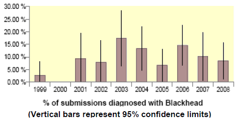
Fewer cases of blackhead were recorded in turkeys during the quarter than last year (Figure 10). However, in one flock 200 out of 700 birds were reported to have died and in another flock 20 out of 600 five-week-old organic turkey poults died and others appeared weak. There were also fewer VIDA records of blackhead in chickens than last year.
(as a percentage of diagnosable submissions)
July – September 1999-2008
Fowl Cholera (Pasteurella multocida)
Three incidents of fowl cholera were recorded on VIDA during the quarter. In one of these swollen faces and a sudden mortality of 20 birds per day over a two day period was described in a group of 2,500 15- week-old turkey poults. Discolouration of the skin of the head with some subcutaneous oedema and swelling of the infra-orbital region along with conjunctivitis was seen. Pasteurella multocida was cultured from sinuses, lungs and airsacs of birds consistent with acute pasteurellosis. The rest of the birds responded well to parenteral penicillin.
Acute septicaemic conditions such pasteurellosis can be confused with avian notifiable diseases including avian influenza.
Marek’s Disease
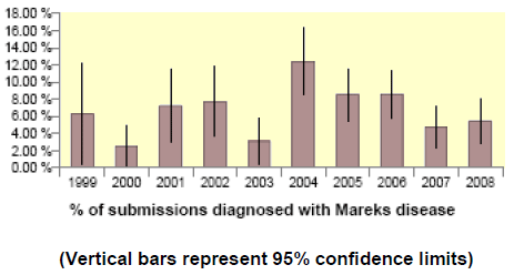
There was a small increase in VIDA diagnoses of Marek’s disease in chickens compared with the same quarter of last year (Figure 11). Most of the recorded incidents were in hobby or backyard chickens, many of which are unvaccinated.
(as a percentage of diagnosable submissions)
July – September 1999-2008
Further Reading
| - | You can view the full report by clicking here. |
December 2008


