US Poultry Industry Manual - response to a highly contagious foreign animal disease
New castle disease and HPAI By:
By: Part of Series:
Editor's Note: The following content is an excerpt from Poultry Industry Manual: The Foreign Animal Disease Preparedness and Response Plan (FAD PReP)/National Animal Health Emergency Management System (NAHEMS) Guidelines which is designed to provide a framework for dealing with an animal health emergency in the United States. Additional content from the manual will be provided as an article series.
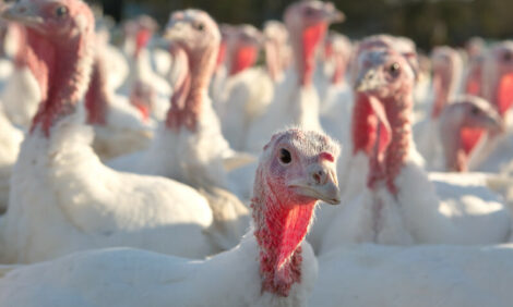
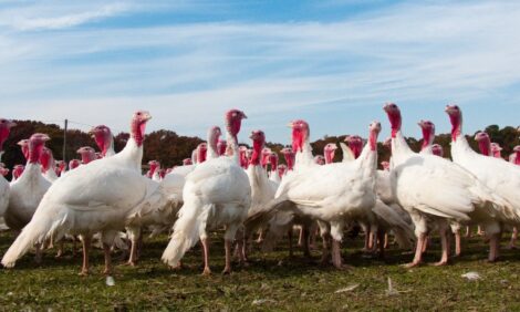
Both Newcastle disease (END) and high pathogenicity avian influenza (HPAI) are nationally and internationally reportable diseases that affect turkeys. In the U.S., infections with low pathogenicity (lentogenic) avian paramyxovirus 1 viruses are still commonly referred to as Newcastle disease, whereas, internationally, infections with these strains are called avian paramyxovirus 1 infections.
Infections of birds with high pathogenicity (mesogenic, velogenic) avian paramyxovirus 1 viruses are termed Exotic Newcastle Disease (END) in the U.S., while internationally these are called just ‘Newcastle Disease’. Strains of low pathogenicity avian influenza (LPAI) viruses that have either H5 or H7 surface hemagglutinins are reportable because they can suddenly mutate from a low to a high pathogenicity virus. Compared to chickens, turkeys are more resistant to ND virus infection but more susceptible to infection with AI viruses. Neither END nor HPAI is endemic in the U.S. so they are referred to as “foreign” or “exotic”. LPAI occurs with some regularity in U.S. commercial and breeder turkey flocks in areas of intense swine production or where there are large numbers of migratory waterfowl. Waterfowl and pigs are frequent sources of AI viruses for turkeys (Capua and Alexander, 2009).
Clinical signs of mild ND or LPAI virus infections are similar and may range from inapparent to a mild respiratory disease characterized by nasal discharge, snicking, rales, dyspnea, conjunctivitis, and sinusitis. Mortality is usually low, but concurrent factors such as age, poor air quality, impaired immunity, and co-infections with bacteria, mycoplasmas, or other viruses can markedly increase mortality. Turkeys typically exhibit decreased activity, huddling, anorexia, and diarrhea. Egg production drops, production of abnormal eggs, and nil to low mortality are seen in affected breeders. Lesions include swollen sinuses, often containing caseous exudate, rhinitis, conjunctivitis, tracheitis, and pulmonary congestion and edema. Swollen kidneys, visceral gout, and multifocal necrosis and hemorrhage in the pancreas also are occasionally seen. Pneumonia, pericarditis, airsacculitis, and splenomegaly are seen when these diseases are complicated by other factors. Yolk peritonitis is commonly seen in breeders, often accompanied by acute oophoritis and salpingitis (Pantin-Jackwood and Swayne, 2009).
Exotic ND in commercial U.S. turkey flocks is extremely rare. However, turkeys are susceptible to experimental infection and become infected when they are commingled with other birds that have the disease (Piacenti, et al. 2006). Clinical signs and gross lesions are similar for END and HPAI.
HPAI is a multi-systemic disease that affects most organs in the body. Early indications of HPAI in both commercial and breeder turkeys are a sudden drop in feed consumption and excessively high, unexplained mortality that may approach 100%. Premonitory clinical signs are usually not observed, but as the disease progresses, turkeys become anorexic, inactive, and non-responsive to stimuli or their environment. Nervous signs characterized by dropped wings, incoordination, tremors, opisthotonos, recumbency, and paddling often occur prior to death. Terminally the birds may flip over and die on their backs. Gross lesions result from necrosis of vascular and lymphoid tissues. Edema, hyperemia, and hemorrhage occur in many organs, but tend to be most obvious in the lungs, proventriculus, ventriculus, pancreas, gut-associated lymphoid tissue, subcutaneous tissues, and fat deposits. Spleens are often enlarged with multifocal necrosis. Similar lesions are seen in breeders along with acute ovarian regression, oophoritis, salpingitis, and yolk peritonitis. There may be no gross lesions in turkeys that die peracutely. Images of poultry affected with HPAI, END, and other diseases can be viewed at http://partnersah.vet.cornell.edu/avian-atlas/.
Early diagnosis and containment are critical for minimizing the impact of an END or HPAI outbreak. All individuals from farm workers to management need to be familiar with the clinical signs and know how to respond if either of these diseases is suspected. Abrupt onset of high mortality with near 100% morbidity is likely to be the earliest indication, especially for HPAI because of the susceptibility of turkeys to avian influenza viruses. A history of LPAI infections in other flocks in the area or unusual mortality among free-living birds, especially waterfowl, may precede an HPAI outbreak. END may not be as obvious, as turkeys are more resistant to infection and vaccination, which is practiced in many turkey producing areas, may modify the response to ND virus infection.
Daily mortality of 0.1% should raise the likelihood of a disease problem, but may not trigger an investigation. Mortality of 1% in any group of turkeys should be investigated to determine the cause. If mortality exceeds this and approaches 10%, an emergency should be declared and the problem investigated immediately. If there is no obvious cause for the deaths (e.g., heat, accident, adverse weather, or poisoning) and there are indications of a possible foreign animal disease, the farm should be shut down and no traffic allowed on or off the farm. The possibility of a foreign animal disease is reported to management while the person investigating the disease stays on the farm to be sure it is secure and waits for a Foreign Animal Disease investigator to arrive. Regulatory authorities, usually the State Veterinarian, are contacted by management. They will send a specialist trained in foreign animal diseases to investigate the problem. It is important that birds not be taken off the farm to a corporate or state diagnostic laboratory. If a disease emergency is declared, and a quarantine established, moving birds to a laboratory will only widen the quarantine zone. Appropriate samples will be collected and submitted to a facility that specializes in diagnosis of foreign animal diseases. Usually, preliminary results to determine if the flock has either END or HPAI are available within 24 hours.
Identification of flocks with LPAI is more problematic as clinical signs are variable and not specific. Often LPAI infection is inapparent and only detected from routine serology for AI done at processing. All flocks submitted for disease diagnosis should be screened for possible LPAI.
Reference: "USDA APHIS | FAD Prep Industry Manuals". Aphis.Usda.Gov. 2013. https://www.aphis.usda.gov/aph...
The manual was produced by the Center for Food Security and Public Health, Iowa State University of Science and Technology, College of Veterinary Medicine, in collaboration with the USDA Animal and Plant Health Inspection Service through a cooperative agreement.

More in this series: Poultry FAD Preparedness & Response
January 2023 - December 2022
 26 January 2023
 5
minute read
5
minute read


 29 December 2022
6
minute read
29 December 2022
6
minute read