


|
Histopathology and Cytology of
Poultry Diseases By Ivan Dinev, DVM, PhD
|
MYCOPLASMA GALLISEPTICUM PNEUMONIA
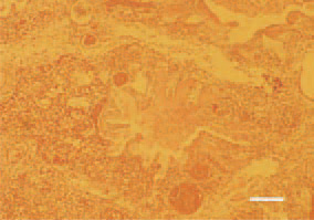
Fig. 1. Congestion and diffuse heterophilic inflitration in the lung parenchyma. The lumen of parabronchi is filled with serous exudate. H/E, Bar = 50 µm.
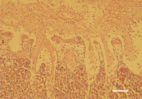
Fig. 2. Croupous pleuropneumonia. Fibrinous pseudomembrane, coating the pleural surface. Cell-rich inflammatory infiltrate among the fibrinous coating and the lung parenchyma. H/E, Bar = 40 µm.
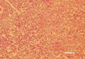
Fig. 3. Airsacculitis, a transverse crosssection of the air sac, broiler chicken. Mixed inflammatory cell exudate and fibrinous caseous masses. H/E, Bar = 35 µm.
This book is protected by the copyright law.
The reproduction, imitation or distribution of the book in whole or in part, in any format (electronic, photocopies etc.) without the prior consent, in writing, of copyright holders is strictly prohibited.