


|
Histopathology and Cytology of
Poultry Diseases By Ivan Dinev, DVM, PhD
|
INCLUSION BODY HEPATITIS

Fig. 1. Cross-section of the liver, 25-day-old broiler chicken. Multiple basophilic intranuclear inclusion bodies on the background of severe degenerative necrobiotic parenchymal lesions. H/E, Bar = 25 µm.
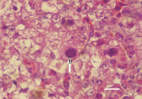
Fig. 2. The basophilic inclusion bodies are usually big and fill tightly the space around the nuclear membrane (arrow – b). H/E, Bar = 10 µm.
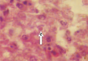
Fig. 3. Eosinophilic inclusion bodies are encountered significantly less frequently, have a round or irregular shape and are surrounded by a light halo (arrow – e). H/E, Bar = 10 µm.
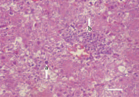
Fig. 4. Although very rarely, along the liver intranuclear inclusion bodies (arrow – a), perivascular mononuclear proliferates (arrow – b) could also be seen. H/E, Bar = 25 µm.
This book is protected by the copyright law.
The reproduction, imitation or distribution of the book in whole or in part, in any format (electronic, photocopies etc.) without the prior consent, in writing, of copyright holders is strictly prohibited.