


|
Histopathology and Cytology of
Poultry Diseases By Ivan Dinev, DVM, PhD
|
COCCIDIOSIS
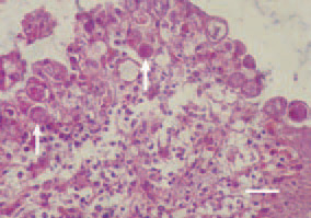
Fig. 1. Histologically, developmental forms (arrows) in a different stage of Eimeria life cycle are detected in epithelial cells of intestines. H/E, Bar = 25 µm.
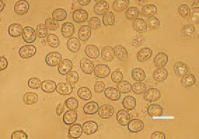
Fig. 2. The microscopic examination of a native preparation of intestinal content or of superficial mucosal layer reveals numerous oocysts in one observation field, native preparation, Bar = 10 µm.
This book is protected by the copyright law.
The reproduction, imitation or distribution of the book in whole or in part, in any format (electronic, photocopies etc.) without the prior consent, in writing, of copyright holders is strictly prohibited.