


|
Histopathology and Cytology of
Poultry Diseases By Ivan Dinev, DVM, PhD
|
INFECTIOUS BURSAL DISEASE (GUMBORO)
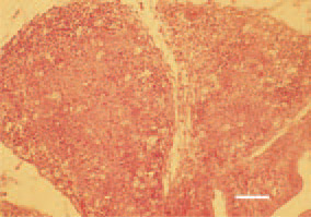
Fig. 1. Almost complete disappearance of the normal follicular structure of B. Fabricii, resulting from severe degenerative necrobiotic lesions and inflammatory cell infiltration. H/E, Bar = 70 µm.
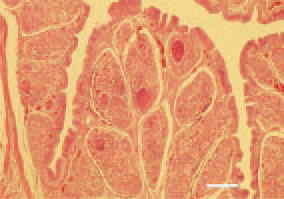
Fig. 2. Marked interfollicular inflammatory oedema, haemorrhages and inflammatory necrotic lesions in the medullary zone of bursal follicles. H/E, Bar = 100 µm.
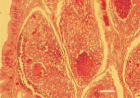
Fig. 3. Fragment of Fig. 2. Inflammatory necrotic detritus among the B. Fabricii follicles. H/E, Bar = 50 µm.
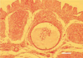
Fig. 4. Sometimes, in the medullary zone of follicles, cystic cavities could be formed that contain exudate, imflammatory cells and detritus mass. H/E, Bar = 35 µm.
This book is protected by the copyright law.
The reproduction, imitation or distribution of the book in whole or in part, in any format (electronic, photocopies etc.) without the prior consent, in writing, of copyright holders is strictly prohibited.