


|
Histopathology and Cytology of
Poultry Diseases By Ivan Dinev, DVM, PhD
|
LYMPHOID LEUKOSIS
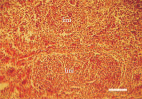
Fig. 1. The neoplastic changes begin always from B. Fabricii, where intrafollicular lymphomas (lim) of a various size are observed. H/E, Bar = 50 µm.
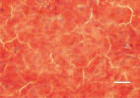
Fig. 2. Histologically, growth of singletype lymphoblast cells with marked pyroninophilia is observed. Tissue cross-section, liver, methyl green pyronin staining, H/E, Bar = 25 µm.
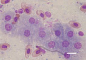
Fig. 3. Equal-sized lymphoblast cells. Touch imprint preparation, liver. Diff Quik, Bar = 10 µm.
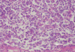
Fig. 4. Lymphoid leukosis, spleen, hen. Growth of large lymphoid cells of a slightly varying size. H/E, Bar = 25 µm.
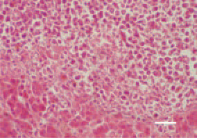
Fig. 5. Liver, lymphoid leukosis, hen. Focal growth of lymphoblasts, single lymphocytes, resulting in atrophy of the parenchyma. H/E, Bar = 25 µm.
This book is protected by the copyright law.
The reproduction, imitation or distribution of the book in whole or in part, in any format (electronic, photocopies etc.) without the prior consent, in writing, of copyright holders is strictly prohibited.