


|
Histopathology and Cytology of
Poultry Diseases By Ivan Dinev, DVM, PhD
|
LARYNGOTRACHEITIS
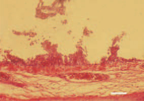
Fig. 1. Trachea, a transverse crosssection. Severe haemorrhagic desquamative inflammation of the mucous coat. Erosions, ulcers and a extremely thinned mucous layer. H/E, Bar = 50 µm.
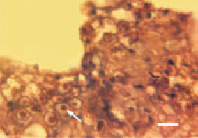
Fig. 2. The detection of eosinophilic intranuclear inclusion bodies (arrow), surrounded by a light halo in the mucous coat epithelium is of essential diagnostic value. This is possible in the initial stage of the disease, prior to the occurrence of desquamative lesions. H/E, Bar = 10 µm.
This book is protected by the copyright law.
The reproduction, imitation or distribution of the book in whole or in part, in any format (electronic, photocopies etc.) without the prior consent, in writing, of copyright holders is strictly prohibited.