


|
Histopathology and Cytology of
Poultry Diseases By Ivan Dinev, DVM, PhD
|
TIBIAL DYSCHONDROPLASIA
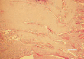
Fig. 1. Longitudinal cross-section, proximal tibia, broiler chicken. Growth and buildup of prehypertrophic cartilage. There is no distinct border between proliferating and hypertrophic cartilage. H/E, Bar = 50 µm.
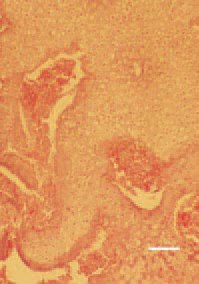
Fig. 2. Flow of abnormal masses of prehypertrophic cartilage, sometimes occupying the entire metaphysis. There is no adequate blood supply, only single blood vessels penetrating from the metaphysis among the abnormal cartilage. H/E, Bar = 35 µm.
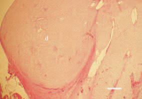
Fig. 3. Subperiosteal dysplasia (d) of prehypertrophic cartilage in the proximal tibia region, resulting in bone deformation. H/E, Bar = 100 µm.
This book is protected by the copyright law.
The reproduction, imitation or distribution of the book in whole or in part, in any format (electronic, photocopies etc.) without the prior consent, in writing, of copyright holders is strictly prohibited.