


|
Diseases of Poultry
By Ivan Dinev, DVM, PhD
|
ESCHERICHIA COLI INFECTIONS
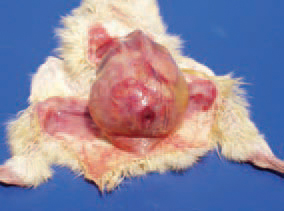
1. Omphalitis (navel infection). It is characterized with reddening and tissue oedema in the umbilical region.
2. Escherichia coli infections are widely distributed among poultry of all ages and categories. They are primarily related to poor hygienic conditions, neglected technological requirements or to respi-ratory and immunosuppressive diseases. A common sequel of navel infections is local or diffuse peritonitis.
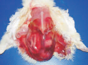
3. When the amount of egg white is bigger (in larger eggs), it impedes the absorption during hatching, resulting in subcutaneous jelly-like oedemas that are an excellent media for the development of E. coli infections.
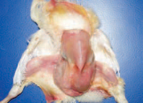
4. The delayed absorption of the yolk sac is a prerequisite for E. coli infections and peritonitis. The most commonly identified E. coli serotypes are: 01:K1 (L); 02:K1 (L) and 078:K80 (B).
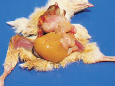
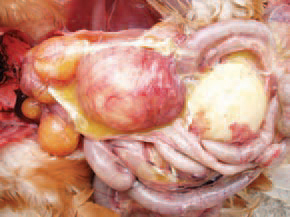
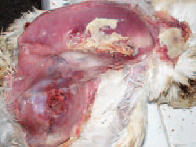
5. At a later stage of the infection, the yolk content is a cause of putrefactive necrotic processes in the peritoneal cavity. The abdomen is bloated. The entire abdominal wall is affected by a moist gangrene (maceration).
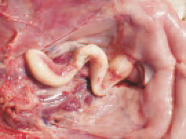
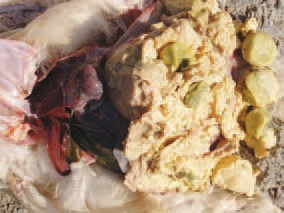
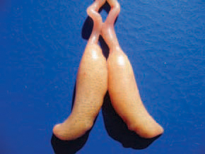
6. Salpingitis (inflammation of the oviduct). Salpingites due to E. coli infections could be also observed in growing birds. The oviduct is dilated, with thinned wall and filled with caseous exudate all along its length.
7. Salpingites are among the commonest causes for death in layer hens. E. coli penetrates from the cloaca via an ascendant route. Predisposing factors are the intense egg laying and the associated estrogen activity.
. 8. Salpingitis. In older cases, the caseous masses in the oviduct have a lamellar structure. E. coli organisms are usually found in excreta because of their presence in avian and mammalian intestine, the birds are constantly at risk of infection through contaminated water, dust, faeces and environment.
9. Salpingitis. Retained yolks among the caseous masses in the oviduct. In some cases, when the systemic resistance is lower, places, contaminated with E. coli, such as intestine, genital tract or nasal passages, could be latent sources of infection.
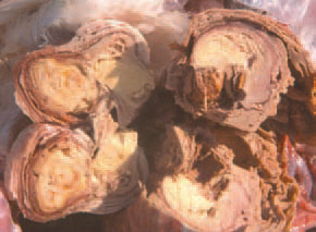
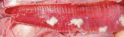
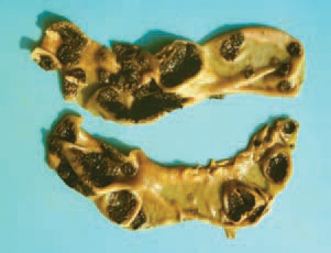
10. Compression and compactedness of caseous necrotic masses after losing a part of their water content in the oviduct of a layer
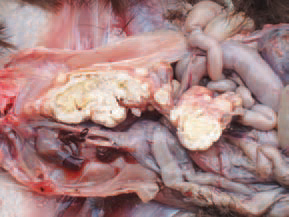
11. Salpingitis. An element of Fig. 10. Longitudinal cross section of the oviduct.
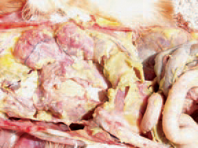
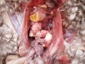
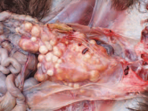
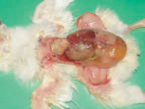
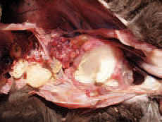
12. Egg yolk peritonitis in a layer hen consequently to E. coli salpingitis. The chickens could be hatched with a latent infection, when E. coli is present in ovaries and the oviduct. In these instances, the infection could turn into an overt infection under the influence of some stress factors or lesions.
13. (inflamation of the ovary) consequently to a salpingitis due to ascendant E coli infection.

14. Cystic degeneration of ovarian follicles following an E. coli opphoritis.
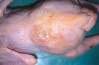
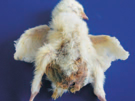
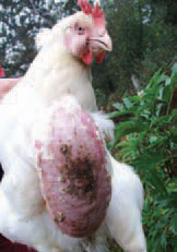
15. Cellulitis (inflammation of the subcutaneous tissue that affects also the overlying skin). It predominates in broilers and is detected mainly in slaughter-houses. Macroscopically, the lesions are with a yellowish-brown colour.
16. Cellulitis. Affected areas are mostly in the region of the back and the thighs.
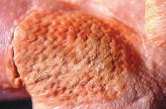
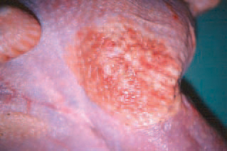
17. In some cases, the lesions are slightly prominating over the adjacent healthy skin.
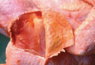
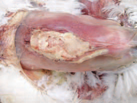
18. Cullulitis. In the subcutaneous tissue, thick fibrinous plaques are often found out.
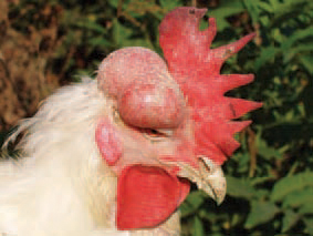
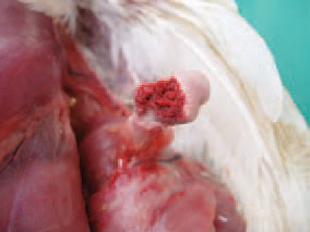
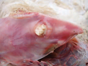
19. In some cases with adult birds, in the region of the head, subcutaneous masses of thick serofibrinous exudate resulting from a local E. coli infection could be detected.
20. Enterocolitis. Enterotoxigenic E. coli that produce toxins, cause the secretion and retention of fluids in some intestinal loops and especially in the caeca. Clinically, diarrhoea and de-hydration are observed. The intestines are pale and distended, particularly the caeca that are overfilled with fluid containing many gas bubbles
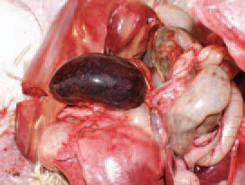
21. . Neonatal E. coli septicaemia. Chickens in the first 24 - 48 h after hatching are affected. The death rate during the first ten days is higher and could reach 5 - 6%. The yolk sac is unabsorbed. The spleen is enlarged. Some days later, the typical serofibrinous polyserositis lesions, affecting the peritoneum, the pericardium, the air sacs and the liver capsule are manifested.
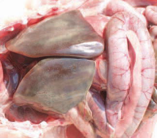
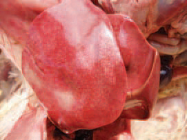
22. Acute E. coli septicaemia in layer hens. Clinically and morphologically, the acute E. coli septicaemia could resemble fowl cholera or fowl typhoid. It is encountered in both young and sexually mature birds. The stress in the beginning of egg-laying is considered as an important predisposing factor. The parenchymal organs are enlarged and hyperaemic. Sometimes, the liver has a greenish colour and is mottled with multiple small necrotic foci. Also, pericarditis, peritonitis and petechial haemorrhages on serous coats are present.
23. 24. 25. E. coli septicaemia of a respiratory origin. In such cases, the respiratory mucosa damaged by infectious and non-infections agents (ND viruses including vaccinal strains, IB, TRT, mycoplasmae, high ammonia levels) is the entrance door of the E. coli infection. The lesions are principally observed in the respiratory tract (trachea, lungs and air sacs), but some adjacent serous coats (pericardium, peritoneum) are also affected and thus, the picture of a typical serofibrinous polyserositis is produced.
26. 27. E. coli septicaemia secondary to enteritis. It is most commonly encountered in turkeys. The intestinal mucosa, damaged by the haemorrhagic enteritis virus (see Adenovirus infections), is the entrance door of E. coli infection. The most typical lesions are the marked enlargement, hyperaemia, haemorrhages and necrosis of the liver and the spleen.
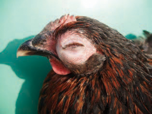
28. Panophthalmitis (inflammation of all tissues of the eyeball). Generally, it develops secondary to E. coli septicaemia and is usually unilateral.
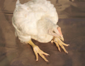
29.. Arthritis, Osteomyelitis and Osteonecrosis (inflammation of joints, bone marrow and bone necrosis, respectively). The lesions are a common sequel to E. coli septicaemia. Clinically, lameness, prolonged lying down, dehydration and retarded growth rate are observed. The coxofemoral joints, the femur and tibiotarsal joints are most commonly affected. The bacteria colonize the physes of growing bones and provoke an inflammatory response that is further causing osteomyelitis. Pathoanatomically, fractures of the femoral head are usually discovered.
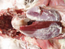
30. 31. some cases of E. coli septicaemia, accumulation of exudate between the superficial and deep pectoral muscles is observed.
32. The lesions that develop in the articular spaces of thoracolumbar vertebrae result in spondylitis (spondylosis) and after that, in progressive paresis and paralysis.
33. Coligranuloma (Hjarre's disease). It is characterized by multiple granulomas in the intestinal tract, the mesentery and the liver, but not in spleen. The lesions are similar to these observed in tuberculosis.
34. 35. . Bursitis sternalis (inflammation of the sternal bursa). The bursa is enlarged in a various extent and filled with inflammatory exudate. The diagnosis of coli - infections is based on isolation and typization of pathogenic E. coli serotypes. Many other bacteria (salmonellae, pasteurellae, staphylococci etc.), viruses, chlamydiae and mycoplasmae should be excluded as possible aetiological agents. The prevention should aim at minimizing the probability of faecal contamination of eggs. This implies the maintenance of clean nests, discarding floor eggs and removal of eggs that are cracked or contaminated with faeces. Breeder eggs should be fumigated or disinfected in the farm prior to their transportation in the storage premise. The treatment is effective if initiated soon after testing the antibacterial sensitivity of isolates.