Book Contents

Diseases of Poultry
By Ivan Dinev, DVM, PhD


VIRAL INCLUSION BODY HEPATITIS

The viral inclusion body hepatitis (IBH) is an adenovirus infection characte-rized by haemorrhages and dystrophic necrobiotic changes in the liver and kidneys, accompanied by intranuclear inclusion bodies. A characte-ristic macroscopic lesion is the enlarged, dystrophic liver with yellowish colour and crumbly texture. The viral inclusion body hepatitis (IBH) is an adenovirus infection characte-rized by haemorrhages and dystrophic necrobiotic changes in the liver and kidneys, accompanied by intranuclear inclusion bodies. A characte-ristic macroscopic lesion is the enlarged, dystrophic liver with yellowish colour and crumbly texture.

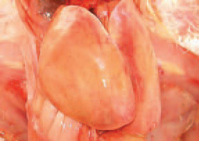
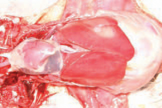
175.176.The viral inclusion body hepatitis (IBH) is an adenovirus infection characte-rized by haemorrhages and dystrophic necrobiotic changes in the liver and kidneys, accompanied by intranuclear inclusion bodies. A characte-ristic macroscopic lesion is the enlarged, dystrophic liver with yellowish colour and crumbly texture.

IBH outbreaks are encountered primarily in meat type chickens, most commonly at the age of 3-8 weeks. IBH often occurs as a secondary infection to immunodeficiency resulting from other diseases (IBD, CIA). On the background of dystrophic liver changes, haemorrhages of various intensity and size are outlined, thus creating a variety of liver lesions. IBH outbreaks are encountered primarily in meat type chickens, most commonly at the age of 3-8 weeks. IBH often occurs as a secondary infection to immunodeficiency resulting from other diseases (IBD, CIA). On the background of dystrophic liver changes, haemorrhages of various intensity and size are outlined, thus creating a variety of liver lesions.

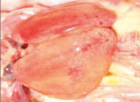
177.178.IBH outbreaks are encountered primarily in meat type chickens, most commonly at the age of 3-8 weeks. IBH often occurs as a secondary infection to immunodeficiency resulting from other diseases (IBD, CIA). On the background of dystrophic liver changes, haemorrhages of various intensity and size are outlined, thus creating a variety of liver lesions.

Throughout IBH outbreaks, several serotypes from the 12 known avian adenoviruses (AAVs) of group I are isolated. The sick chickens cany the virus in their excreta, kidneys, tracheal and nasal mucosa. The virus is resistant to many environmental factors and could be easily trans-mitted by a mechanical route. The transmission of adenoviruses is realized vertically by breeder eggs and horizontally, via excreta (mainly faeces). In a number of cases, the dominating lesion is the massive mottled or striated haemorrhages of the liver. Throughout IBH outbreaks, several serotypes from the 12 known avian adenoviruses (AAVs) of group I are isolated. The sick chickens cany the virus in their excreta, kidneys, tracheal and nasal mucosa. The virus is resistant to many environmental factors and could be easily trans-mitted by a mechanical route. The transmission of adenoviruses is realized vertically by breeder eggs and horizontally, via excreta (mainly faeces). In a number of cases, the dominating lesion is the massive mottled or striated haemorrhages of the liver.

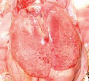
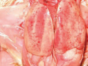
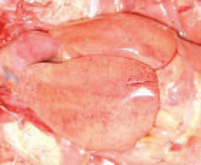
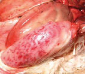
179.180.Throughout IBH outbreaks, several serotypes from the 12 known avian adenoviruses (AAVs) of group I are isolated. The sick chickens cany the virus in their excreta, kidneys, tracheal and nasal mucosa. The virus is resistant to many environmental factors and could be easily trans-mitted by a mechanical route. The transmission of adenoviruses is realized vertically by breeder eggs and horizontally, via excreta (mainly faeces). In a number of cases, the dominating lesion is the massive mottled or striated haemorrhages of the liver.

IBH is characterized by a sudden onset and a sharply increased death rate that reaches peak values by the 3rd - 4th day and returns back within the normal range by the 6th - 7th day. The total death rate is usually under 10% but sometimes could attain 30%. More rarely, macro-scopically visible necrotic foci could be detected in the liver.

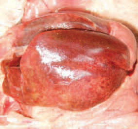
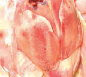
181.IBH is characterized by a sudden onset and a sharply increased death rate that reaches peak values by the 3rd - 4th day and returns back within the normal range by the 6th - 7th day. The total death rate is usually under 10% but sometimes could attain 30%. More rarely, macro-scopically visible necrotic foci could be detected in the liver.

The kidneys are enlarged, pale and mottled with multiple haemorrhages.

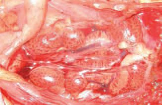
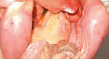
182.The kidneys are enlarged, pale and mottled with multiple haemorrhages.

many instances of IBH, the amount of pericardial fluid is increased  (hydropericardium).

183.many instances of IBH, the amount of pericardial fluid is increased (hydropericardium).

Clinical signs could be observed only several hours prior to death occurrence. They consist in pale comb and wattles, depression and apathy. Sometimes, the skin is icteric. Often, ecchymoses and striated haemorrhages in skeletal muscles are observed. Clinical signs could be observed only several hours prior to death occurrence. They consist in pale comb and wattles, depression and apathy. Sometimes, the skin is icteric. Often, ecchymoses and striated haemorrhages in skeletal muscles are observed.

184.185.Clinical signs could be observed only several hours prior to death occurrence. They consist in pale comb and wattles, depression and apathy. Sometimes, the skin is icteric. Often, ecchymoses and striated haemorrhages in skeletal muscles are observed.

Microscopically, extensive dystrophic changes and necroses of liver parenchyma are detected. In the nuclei of hepatocytes, basophilic or eosinophilic inclusion bodies are detected. The basophilic inclusion bodies are usually dense and occupy the entire nuclear inner space (arrow b), whereas the eosinophilic ones are round or irregularly shaped and surrounded by a light halo (arrow e). The diagnosis is based upon the typical gross lesions and the history records. Microscopically, extensive dystrophic changes and necroses of liver parenchyma are detected. In the nuclei of hepatocytes, basophilic or eosinophilic inclusion bodies are detected. The basophilic inclusion bodies are usually dense and occupy the entire nuclear inner space (arrow b), whereas the eosinophilic ones are round or irregularly shaped and surrounded by a light halo (arrow e). The diagnosis is based upon the typical gross lesions and the history records.

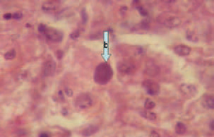
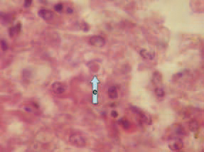
186\186"Microscopically, extensive dystrophic changes and necroses of liver parenchyma are detected. In the nuclei of hepatocytes, basophilic or eosinophilic inclusion bodies are detected. The basophilic inclusion bodies are usually dense and occupy the entire nuclear inner space (arrow b), whereas the eosinophilic ones are round or irregularly shaped and surrounded by a light halo (arrow e). The diagnosis is based upon the typical gross lesions and the history records.A principal approach in IBH diagnostics is the histological investigation that helps to detect the intranuclear inclusion bodies. IBH should be distinguished mainly from IBD and chicken infectious anaemia (CIA). With regard to IBH prevention and control, the eggs of broiler parent flocks, where the disease is consecutively appearing in the progeny, should not be used for hatching. The access of wild birds should be prevented as they are potential carriers and distributors of the virus. The most important steps in IBH prevention are the control of IBD and CIA. There are neither vaccines, nor an effective treatment.

This book is protected by the copyright law. The reproduction, imitation or distribution of the book in whole or in part, in any format (electronic, photocopies etc.) without the prior consent, in writing, of copyright holders is strictly prohibited.
© 2000 - 2024 - Global Ag Media. All Rights Reserved | No part of this site may be reproduced without permission.