


|
Diseases of Poultry
By Ivan Dinev, DVM, PhD
|
MYELOCYTOMATOSIS
311.312.MC is caused by viral staings of ALSVs from sungroups A,B and J (Mc29, MC31, Cm11, OK10, HRPS 103, and ADOL HC1). It is encountered relatively infrequently. Its occurrence is sporadic or enzootic. Suceptible birds and hens, pheasents, guinea hens and quails. In most cases, the liver is enlarged, thick and mottled with dark red sports or fat like nodules.
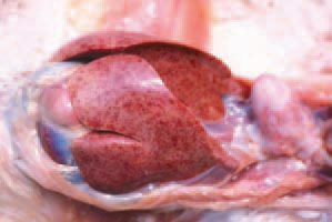
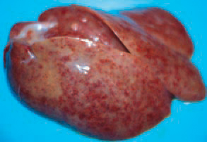
313.314.. MC is caused by viral strains of ALSVs from subgroups A, B and J (MC29, MC31, CMII, OK10, HRPS 103, and ADOL HC1). It is encountered relatively infrequently. Its occurrence is sporadic or enzootic. Susceptible birds are hens, pheasants, guinea hens and quails. In most cases, the liver is enlarged, thick and mottled with dark red spots or fat-like nodules.
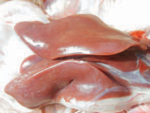
315.Sclerotic changes in the liver are possible because of regression of neoplastic lesions.
316.The spleen is usually enlarged, but sometimes, could be atrophied.
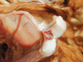
317.. A characteristic feature of MC is its simultaneous course with tumours from a different type: mesenchymal, epithelial or mixed. The picture shows a fibrosarcoma to the gizzard associated with MC.
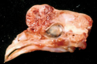
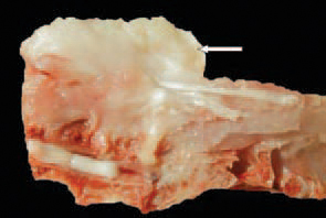
318.Mixed mesenchymal tumour (osteochondrosarcoma) to the frontal skull bones: a sagittal cross section
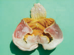
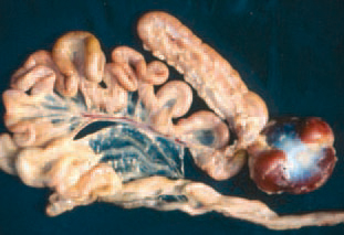
319.Multiple rabdomyosarcoma in pectoral, thigh, abdominal and tracheal muscles.
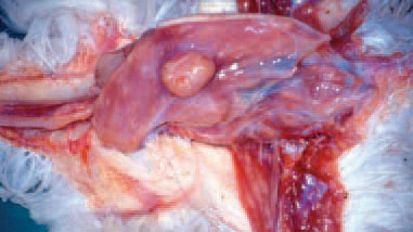
320.Leiomyosarcoma of the mucous coat on the oviduct.
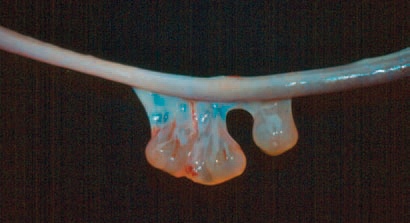
321.Pendulation harmangio-sarcoma of the ileal serosa.
322.Pendulating multiple myxoma of the small intestine's serous coat.
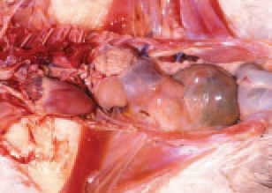
323.. MC-associated cystadenocarcinoma of the kidney in a hen.
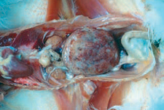
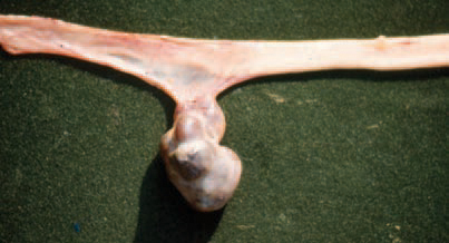
324.Nephroblastoma of the left kidney, occupying a significant part of the abdominal cavity.
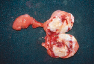
325.Nephroblastoma -the surface of a cross section. The tumour is a pendulating mass attached to the kidney by a fibrous vascularized stem that has undergone a partial necrosis and haemorrhages
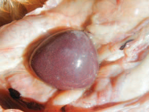
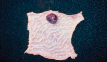
326.Granulosa cell tumour of the ovary. The tumour appears as a single, compact, dorsoventrally flattened growth.
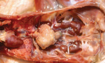
327.MC-associated multiple carcino-sarcoma of the mesentery and alimentary tract's serous coat (disseminated milliary nodules).
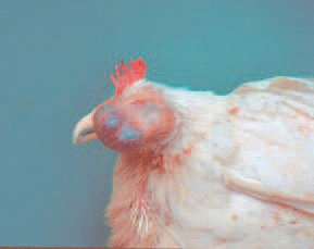
328.MC-associated carcinosarcomas in the region of the right infraorbital sinus.
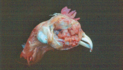
329.Gross appearance of the tumour from Fig. 328 after removal of the covering skin.
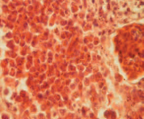
330.Histologically, myelocytomatomas are easily distinguished. Most commonly, they have perivascular localization. Growth of myelocytes with well-formed granules in a liver cross-section.
331.Kidney.Focal intertubular myelocytic pro-liferations.
332.. MC-associated neoplasms of epithelial, mesenchymal or mixed type demonstrate the respective type of histological structure. Leiomyosarcoma a histological view. Polygonal giant cells with hyperchromatic nuclei.
333.Leiomyosarcoma - small intestime. Prolongations of polynuclear symplastic elements.
334.Leiomyosarcoma - small intestine. Extraordinary („monstrous") multinuclear giant cell with intracytoplasmic vacuoles.
335.. Rabdomyosarcoma. An area with multiple hyperchromatic giant cells.
336.Carcinosarcoma of the pancreas. Tubulous glandular epithelial formations of the carcinoma component among the liposarcoma part of the parenchyma. The diagnosis is based upon the entity of data about the history, the gross appearance and location of the tumours and the specific histo-logical lesions. From a differential diagnostic point of view, myelo-blastosis and erythroblastosis should be considered.