Book Contents

Diseases of Poultry
By Ivan Dinev, DVM, PhD


CHOLANGIOHEPATITIS IN BROILER CHICKENS

Cholangiohepatitis (CAH) in broiler chickens is characterized by inflammatory proliferative and dystrophic necrobiotic alterations in bile ducts and the liver parenchyma. Usually, no clinical signs are observed. The increased daily mortality is insignificant, although in some chickens, a retarded growth and dehydration could be present. Pathoanatomically, the liver is enlarged and with paler yellow colour. In some cases, its surface has a characteristic acinous appearance and in others is mottled with multiple small greyish-white or greenish foci.

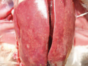
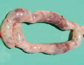
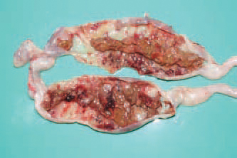
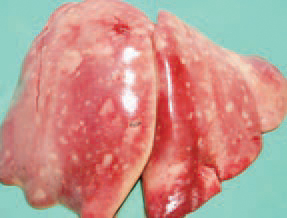
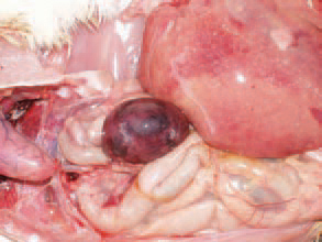
125.126.Cholangiohepatitis (CAH) in broiler chickens is characterized by inflammatory proliferative and dystrophic necrobiotic alterations in bile ducts and the liver parenchyma. Usually, no clinical signs are observed. The increased daily mortality is insignificant, although in some chickens, a retarded growth and dehydration could be present. Pathoanatomically, the liver is enlarged and with paler yellow colour. In some cases, its surface has a characteristic acinous appearance and in others is mottled with multiple small greyish-white or greenish foci.

The walls of the gall bladder are thickened, sometimes up to 5-6 cm, and opaque. The state is detected in the last phase of the fattening period or in the slaughterhouse. It is possible to observe CHA as an independent disease or associated with necrotic enteritis.

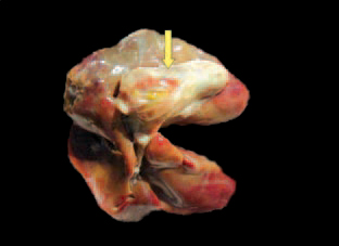
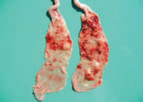
127The walls of the gall bladder are thickened, sometimes up to 5-6 cm, and opaque. The state is detected in the last phase of the fattening period or in the slaughterhouse. It is possible to observe CHA as an independent disease or associated with necrotic enteritis.

A transverse cross section through the thickened gall bladder wall

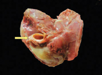
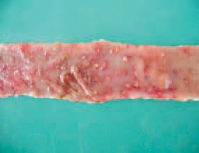
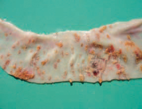
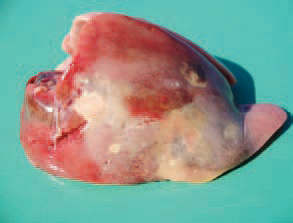
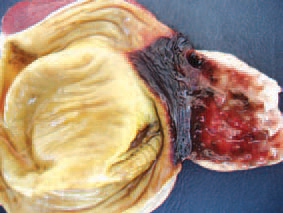
128A transverse cross section through the thickened gall bladder wall

Clostridium perfringens is the aetiological agent. CAH is experimentally reproduced in broilers by ligation of bile ducts and inoculation with CI. perfringens. The gall bladder is filled with a thick bile secretion or a dense matter with a creamy colour.

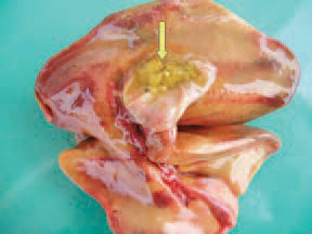
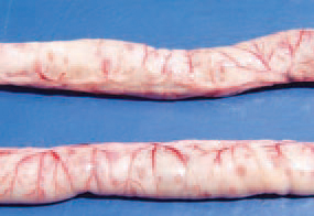
129Clostridium perfringens is the aetiological agent. CAH is experimentally reproduced in broilers by ligation of bile ducts and inoculation with CI. perfringens. The gall bladder is filled with a thick bile secretion or a dense matter with a creamy colour.

In some chickens, the sub-cutaneous fat and the body fat have an icteric tint.

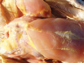
130In some chickens, the sub-cutaneous fat and the body fat have an icteric tint.

Histologically, the liver lesions are detected at a various stage of development. In the majority of cases, proliferative changes in bile ducts are observed. The overgrown bile ducts form granulomatous struc-tures, surrounded by fine reticular fibres. Centrally, in some granulomas, either initial or advanced degree of necrosis and weak to moderate granulocytic infiltration are observed.

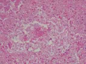
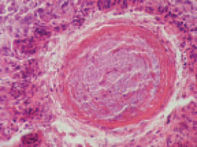
131.Histologically, the liver lesions are detected at a various stage of development. In the majority of cases, proliferative changes in bile ducts are observed. The overgrown bile ducts form granulomatous struc-tures, surrounded by fine reticular fibres. Centrally, in some granulomas, either initial or advanced degree of necrosis and weak to moderate granulocytic infiltration are observed.

In many bile ducts, a biliary stasis is present and within some, among the stagnated secretion, a huge amount of microorganisms are detected. Pericanalicularly, coagulation necroses are frequently noticed. Among some of these foci, hyalinization of the necrotic masses and single microorganisms are present. Around the necroses, a belt of macrophages, lymphocytes and granulocytes is formed.

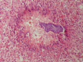
132In many bile ducts, a biliary stasis is present and within some, among the stagnated secretion, a huge amount of microorganisms are detected. Pericanalicularly, coagulation necroses are frequently noticed. Among some of these foci, hyalinization of the necrotic masses and single microorganisms are present. Around the necroses, a belt of macrophages, lymphocytes and granulocytes is formed.

. Many Gram-positive bacteria are detected among granulomas, in bile ducts' lumen and into the gall bladder, often accompanied by inflammatory lesions. The mucosa of bile ducts and the gall bladder is frequently necrotized and the wall is thickened because of connective tissue growth. The diagnosis is based on the characteristic gross and microscopic lesions. The prevention is similar to that in NE.

133. Many Gram-positive bacteria are detected among granulomas, in bile ducts' lumen and into the gall bladder, often accompanied by inflammatory lesions. The mucosa of bile ducts and the gall bladder is frequently necrotized and the wall is thickened because of connective tissue growth. The diagnosis is based on the characteristic gross and microscopic lesions. The prevention is similar to that in NE.

Ulcerative enteritis (UE) is characterized by inflammatory ulcerative and necrotic changes in intestinal mucosa and dystrophic necrobiotic lesions of the liver and the spleen. The clinical signs include a general malaise, ruffled feathers, diarrhoea and anaemia. In many instances, the disease begins with a sudden death. Pathoanatomically, deep button-like ulcers are observed, mainly in caeca and less frequently, in some parts of the small intestine, usually visible through the wall. Ulcerative enteritis (UE) is characterized by inflammatory ulcerative and necrotic changes in intestinal mucosa and dystrophic necrobiotic lesions of the liver and the spleen. The clinical signs include a general malaise, ruffled feathers, diarrhoea and anaemia. In many instances, the disease begins with a sudden death. Pathoanatomically, deep button-like ulcers are observed, mainly in caeca and less frequently, in some parts of the small intestine, usually visible through the wall.

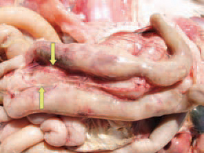
134. 135.Ulcerative enteritis (UE) is characterized by inflammatory ulcerative and necrotic changes in intestinal mucosa and dystrophic necrobiotic lesions of the liver and the spleen. The clinical signs include a general malaise, ruffled feathers, diarrhoea and anaemia. In many instances, the disease begins with a sudden death. Pathoanatomically, deep button-like ulcers are observed, mainly in caeca and less frequently, in some parts of the small intestine, usually visible through the wall.

UE is a problem in all world regions with extensive poultry breeding. Young birds are infected more frequently although the disease is also common among adult quails. The early lesions appear like yellowish foci with haemorrhagic boundaries that could be seen from both the serous and the mucosal surfaces. UE is a problem in all world regions with extensive poultry breeding. Young birds are infected more frequently although the disease is also common among adult quails. The early lesions appear like yellowish foci with haemorrhagic boundaries that could be seen from both the serous and the mucosal surfaces.

136.137.UE is a problem in all world regions with extensive poultry breeding. Young birds are infected more frequently although the disease is also common among adult quails. The early lesions appear like yellowish foci with haemorrhagic boundaries that could be seen from both the serous and the mucosal surfaces.

The aetiological agent is Clostridium colinum, a spore-forming organism, highly resistant to chemical agents and physical alterations. The intestinal content is often mixed with blood.

138.The aetiological agent is Clostridium colinum, a spore-forming organism, highly resistant to chemical agents and physical alterations. The intestinal content is often mixed with blood.

In older and larger ulcers, the haemorrhagic zones tend to disappear. The ulcers could have an irregular round or elongated shape and are covered by large necrotic diphtheritic membranes.

139.In older and larger ulcers, the haemorrhagic zones tend to disappear. The ulcers could have an irregular round or elongated shape and are covered by large necrotic diphtheritic membranes.

Frequently, adhesive peritonitis due to inflammatory involvement of adjacent serous coats is observed. Numerous domestic and wild birds (chickens, quails, turkeys, rock partridges, geese, partridges etc.) are susceptible. The chickens and the quails are the most vulnerable between 4 and 12 weeks of age where as turkeys between 3 and 8 weeks of age.

140.Frequently, adhesive peritonitis due to inflammatory involvement of adjacent serous coats is observed. Numerous domestic and wild birds (chickens, quails, turkeys, rock partridges, geese, partridges etc.) are susceptible. The chickens and the quails are the most vulnerable between 4 and 12 weeks of age where as turkeys between 3 and 8 weeks of age.

liver, a variety of dystrophic changes and necroses with different size and shape are detected. Necrotic foci in some cases are milliary.

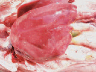
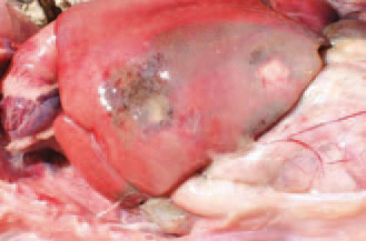
141.liver, a variety of dystrophic changes and necroses with different size and shape are detected. Necrotic foci in some cases are milliary.

Sometimes, liver necroses reach 1 - 2 cm. in diameter and are surrounded by a haemorrhagic zone.

142.Sometimes, liver necroses reach 1 - 2 cm. in diameter and are surrounded by a haemorrhagic zone.

In some instances, necrotic foci involve large parts of the liver and are infiltrated by haemorrhages. The aetiolo-gical agent is distributed with the excreta of acutely ill and recovered birds and persists in soil for many months. The incubation period is 1 - 3 days. The death rate in chickens varies from 2% to 10% and in quails reaches 100%. The outbreaks of UE in chickens are often associated with or come after coccidioses, CIA, IBD or stress conditions. In some instances, necrotic foci involve large parts of the liver and are infiltrated by haemorrhages. The aetiolo-gical agent is distributed with the excreta of acutely ill and recovered birds and persists in soil for many months. The incubation period is 1 - 3 days. The death rate in chickens varies from 2% to 10% and in quails reaches 100%. The outbreaks of UE in chickens are often associated with or come after coccidioses, CIA, IBD or stress conditions.

143.144.In some instances, necrotic foci involve large parts of the liver and are infiltrated by haemorrhages. The aetiolo-gical agent is distributed with the excreta of acutely ill and recovered birds and persists in soil for many months. The incubation period is 1 - 3 days. The death rate in chickens varies from 2% to 10% and in quails reaches 100%. The outbreaks of UE in chickens are often associated with or come after coccidioses, CIA, IBD or stress conditions.

. Most commonly, necroses are distinguished on the background of a marked parenchymatous dystrophy, affecting partially or totally (uni- or bilaterally) the liver.

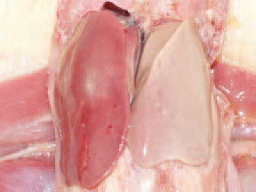
145.. Most commonly, necroses are distinguished on the background of a marked parenchymatous dystrophy, affecting partially or totally (uni- or bilaterally) the liver.

The spleen could be enlarged, hyperaemic, haemorrhagic and some-times, with necroses. The diagnosis is based on the typical gross lesions. When needed, imprint preparations are made, a histological study is performed or attempts for isolation and identification of the aetiological agent are made. UE should be differentiated from NE, coccidiosis and histomonosis (typhlo-hepatitis).

146.The spleen could be enlarged, hyperaemic, haemorrhagic and some-times, with necroses. The diagnosis is based on the typical gross lesions. When needed, imprint preparations are made, a histological study is performed or attempts for isolation and identification of the aetiological agent are made. UE should be differentiated from NE, coccidiosis and histomonosis (typhlo-hepatitis).

some cases, haemorrhages with various intensities are detected in the mucous coat of the gizzard. Prevention -separate housing of the different age groups of birds, avoiding the contact with other avian species. The pre-medication of forages with some antibiotics and their rotation would prevent the reproduction of CI. colinum. A good effect is achieved with oxytetra-cycline dihydrate (OTC 50% premix). UE could be effectively treated with doxycycline hydrochloride, amoxycillin etc.

147.some cases, haemorrhages with various intensities are detected in the mucous coat of the gizzard. Prevention -separate housing of the different age groups of birds, avoiding the contact with other avian species. The pre-medication of forages with some antibiotics and their rotation would prevent the reproduction of CI. colinum. A good effect is achieved with oxytetra-cycline dihydrate (OTC 50% premix). UE could be effectively treated with doxycycline hydrochloride, amoxycillin etc.

This book is protected by the copyright law. The reproduction, imitation or distribution of the book in whole or in part, in any format (electronic, photocopies etc.) without the prior consent, in writing, of copyright holders is strictly prohibited.
© 2000 - 2024 - Global Ag Media. All Rights Reserved | No part of this site may be reproduced without permission.