Diseases of Poultry
By Ivan Dinev, DVM, PhD
|
ASPERGILLUS GRANULOMATOUS DERMATITIS
![389](https://cdn.globalagmedia.com/poultry/legacy/publications/images/image_Page_073_Image_0004.jpg)
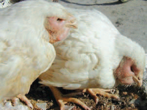
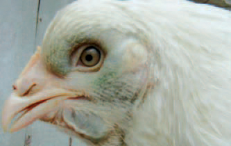
389.The Aspergillus granulomatous dermatitis as a postvaccinal compli-cation is observed in growing broiler breeders. Following subcutaneous application of oil adjuvant vaccine in the lower part of the neck, a marked local reaction with extensive swelling of the head is observed.
![390](https://cdn.globalagmedia.com/poultry/legacy/publications/images/image_Page_073_Image_0006.jpg)
![391](https://cdn.globalagmedia.com/poultry/legacy/publications/images/image_Page_073_Image_0005.jpg)
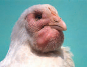
390.391.. The swellings involve the subcutaneous tissue of periorbital sinuses, the mandibular space, even the entire skull without the skin adnexa.
![The head is](https://cdn.globalagmedia.com/poultry/legacy/publications/images/image_Page_074_Image_0001.jpg)
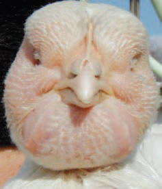
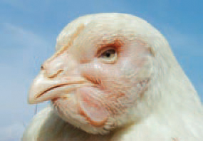
392.The head is ballooned with strongly stretched skin.
![in some cases](https://cdn.globalagmedia.com/poultry/legacy/publications/images/image_Page_074_Image_0002.jpg)
393.In some cases, the covering skin acquires a blue-greenish colour.
![The eye](https://cdn.globalagmedia.com/poultry/legacy/publications/images/image_Page_074_Image_0003.jpg)
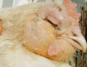
394.The eyeball is sometimes affected by necrotic colliquation.
![Among the cross sectional surface if the highly oedematous subcutaneous tissue, multiple structures resembling sand grains are detected.](https://cdn.globalagmedia.com/poultry/legacy/publications/images/image_Page_074_Image_0004.jpg)
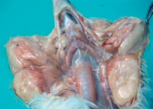
395.Among the cross sectional surface if the highly oedematous subcutaneous tissue, multiple structures resembling sand grains are detected.
![a later stage, a spontaneous regression of oedemas occurs, but granulomatous formations continue to be visible in the subcutaneous tissue.](https://cdn.globalagmedia.com/poultry/legacy/publications/images/image_Page_074_Image_0005.jpg)
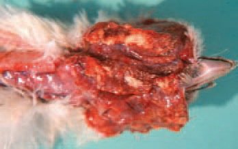
396.a later stage, a spontaneous regression of oedemas occurs, but granulomatous formations continue to be visible in the subcutaneous tissue.
![Histologically, among the oedematous subcutaneous connective tissue, multiple granulomas are detected.](https://cdn.globalagmedia.com/poultry/legacy/publications/images/image_Page_074_Image_0006.jpg)
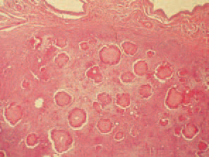
397.Histologically, among the oedematous subcutaneous connective tissue, multiple granulomas are detected.
![central zone of granulomas is an inflammatory necrotic detritus with many eosinophilic leukocytes delineated within.](https://cdn.globalagmedia.com/poultry/legacy/publications/images/image_Page_075_Image_0001.jpg)
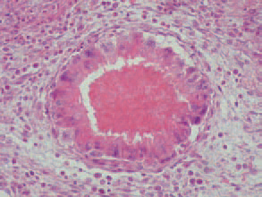
398.central zone of granulomas is an inflammatory necrotic detritus with many eosinophilic leukocytes delineated within.
![, as a crown, multinuclear giant cells of a foreign-body type are arranged, with fibroblast fibrocyte growths around them.](https://cdn.globalagmedia.com/poultry/legacy/publications/images/image_Page_075_Image_0002.jpg)
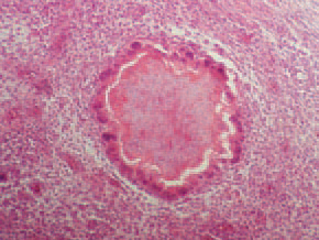
399., as a crown, multinuclear giant cells of a foreign-body type are arranged, with fibroblast fibrocyte growths around them.
![. At a later stage of the condition, after phagocytosis of the detritus, the structure of granulomas is mainly composed by foreign-body giant cells and connective tissue growths.](https://cdn.globalagmedia.com/poultry/legacy/publications/images/image_Page_075_Image_0003.jpg)
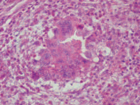
400.. At a later stage of the condition, after phagocytosis of the detritus, the structure of granulomas is mainly composed by foreign-body giant cells and connective tissue growths.